Umbilical cord blood mononuclear cell separation method and application thereof
A separation method and nuclear cell technology, applied in the direction of cell dissociation method, blood/immune system cells, animal cells, etc., can solve the problems of high technical requirements and complicated operation, and achieve the reduction of technical requirements, stable separation effect, and time-consuming short effect
- Summary
- Abstract
- Description
- Claims
- Application Information
AI Technical Summary
Problems solved by technology
Method used
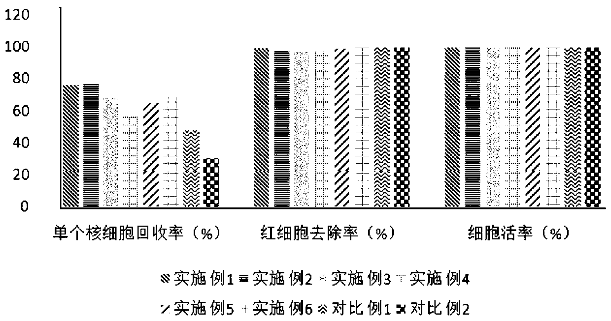
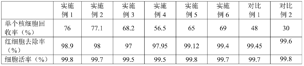
Image

Examples
Embodiment 1
[0027] 1. Add 20mL Ficoll to a 50mL centrifuge tube (recorded as tube ①).
[0028] 2. Add 20mL of umbilical cord blood into a 50mL centrifuge tube (denoted as tube ②), shake well, and perform cell counting. The mononuclear cells in the whole blood are 13.1×10 9 pc / L, whole red blood cell is 3.42×10 12 a / L.
[0029] 3. Use a pipette to directly inject the umbilical cord blood in tube ② into tube ① containing Ficoll, mix it upside down, and let it stand for processing.
[0030] 4. After standing still for 40 minutes, it can be seen that the tube is divided into 3 layers, the upper layer is plasma, the lower layer is red blood cells and granulocytes, and the middle layer is buffy coat mainly composed of mononuclear cells.
[0031] 5. Aspirate the buffy coat with a pipette, transfer the buffy coat into a 50mL centrifuge tube (denoted as tube ③), wash it twice with normal saline, and centrifuge at 800g for 8 minutes each time.
[0032] 6. Finally, resuspend the cells with 10ml o...
Embodiment 2
[0034] 1. Add 30mL Ficoll to a 50mL centrifuge tube (marked as tube ①).
[0035] 2. Add 20mL of umbilical cord blood into a 50mL centrifuge tube (denoted as tube ②), shake well, and perform cell counting. The mononuclear cells in the whole blood are 7.5×10 9 pc / L, whole blood red blood cell is 3.18×10 12 a / L.
[0036] 3. Use a pipette to directly inject the umbilical cord blood in tube ② into tube ① containing Ficoll, mix it upside down, and let it stand for processing.
[0037] 4. After standing still for 40 minutes, it can be seen that the tube is divided into 3 layers, the upper layer is plasma, the lower layer is red blood cells and granulocytes, and the middle layer is buffy coat mainly composed of mononuclear cells.
[0038] 5. Aspirate the buffy coat with a pipette, transfer the buffy coat into a 50mL centrifuge tube (denoted as tube ③), wash it twice with normal saline, and centrifuge at 800g for 8 minutes each time.
[0039] 6. Finally, resuspend the cells with 10m...
Embodiment 3
[0041] 1. Add 40mL Ficoll to a 250mL centrifuge tube (recorded as tube ①).
[0042] 2. Add 20mL of umbilical cord blood into a 250mL centrifuge tube (denoted as tube ②), shake well, and perform cell counting. The mononuclear cells in the whole blood are 9.8×10 9 pc / L, whole blood red blood cell is 3.16×10 12 a / L.
[0043] 3. Use a pipette to directly inject the umbilical cord blood in tube ② into tube ① containing Ficoll, mix it upside down, and let it stand for processing.
[0044] 4. After standing still for 40 minutes, it can be seen that the tube is divided into 3 layers, the upper layer is plasma, the lower layer is red blood cells and granulocytes, and the middle layer is buffy coat mainly composed of mononuclear cells.
[0045] 5. Aspirate the buffy coat with a pipette, transfer the buffy coat into a 50mL centrifuge tube (denoted as tube ③), wash it twice with normal saline, and centrifuge at 800g for 8 minutes each time.
[0046] 6. Finally, resuspend the cells with...
PUM
 Abstract
Description
Claims
Application Information
 - R&D Engineer
- R&D Manager
- IP Professional
- Industry Leading Data Capabilities
- Powerful AI technology
- Patent DNA Extraction
Browse by: Latest US Patents, China's latest patents, Technical Efficacy Thesaurus, Application Domain, Technology Topic.
© 2024 PatSnap. All rights reserved.Legal|Privacy policy|Modern Slavery Act Transparency Statement|Sitemap