Lung pathological image classification and segmentation method based on deep learning

A pathological image and deep learning technology, applied in the field of medical image analysis, to achieve the effect of solving the inconsistency of segmentation results

Pending Publication Date: 2020-10-16
MOTIC XIAMEN MEDICAL DIAGNOSTICS SYST
View PDF0 Cites 5 Cited by
  • Summary
  • Abstract
  • Description
  • Claims
  • Application Information

AI Technical Summary

Problems solved by technology

[0006] Aiming at the defects of the existing classification and segmentation of lung pathological images, the present invention proposes a method for classification and segmentation of lung pathological images based on deep learning

Method used


Image

  • Lung pathological image classification and segmentation method based on deep learning
  • Lung pathological image classification and segmentation method based on deep learning
  • Lung pathological image classification and segmentation method based on deep learning

Examples


Embodiment Construction

[0043] The present invention will be further described below in conjunction with the drawings. The following embodiments are only used to explain the technical solutions of the present invention more clearly, and cannot be used to limit the protection scope of the present invention.

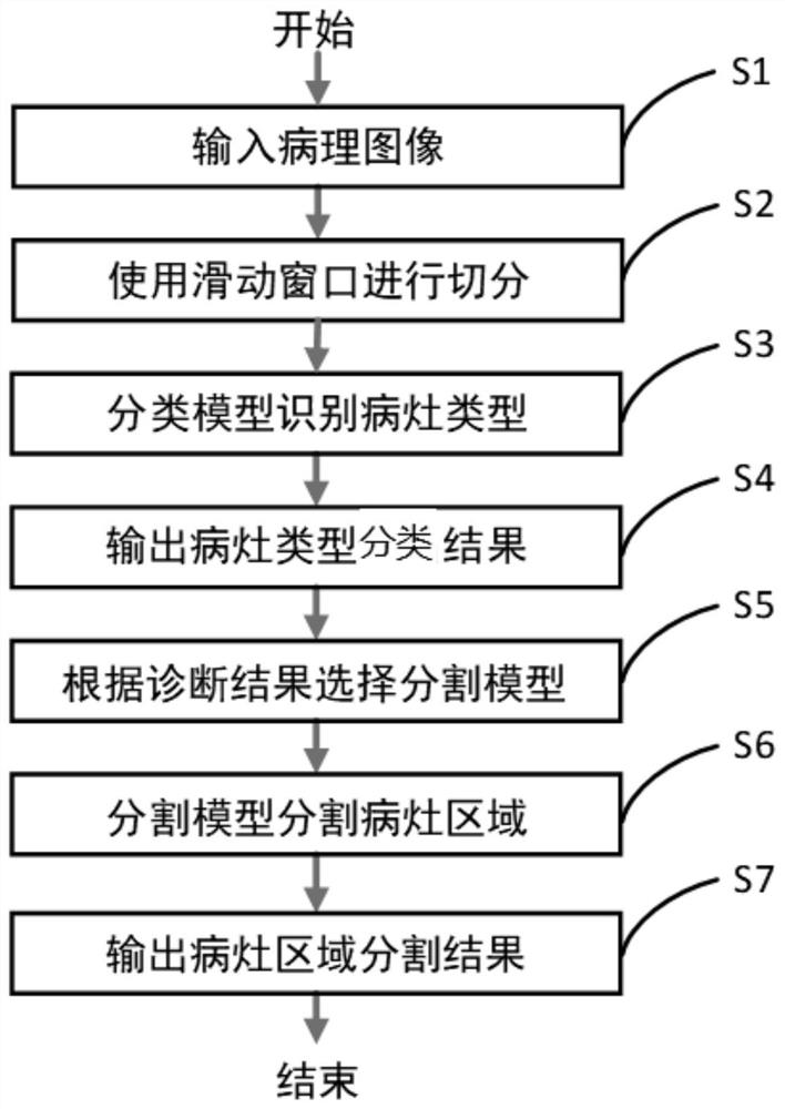
[0044] Such as figure 1 As shown, a method for classification and segmentation of lung pathological images based on deep learning provided by the present invention includes the following steps:

[0045] Step S1: Input the pathological image of the whole slice;

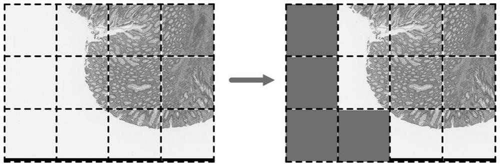
[0046] Step S2: Use a sliding window to segment the pathological image to obtain image blocks;

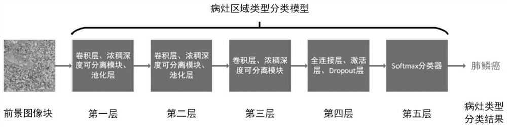
[0047] Step S3: Use the lesion type classification model to sequentially analyze the foreground image blocks, and identify the lesion type in the tissue area in the foreground image block;

[0048] Step S4: Output the classification result of the lesion type;

[0049] Step S5: Select a corresponding lesion area segmentation model according to the classi...


PUM


Abstract

The invention relates to the technical field of medical image analysis, in particular to a lung pathological image classification and segmentation method. The method comprises the following steps: inputting a pathological image of a full slice; segmenting the pathological image by using a sliding window to obtain image blocks; analyzing foreground image blocks in sequence by using a focus type classification model, and identifying focus types of tissue regions in the foreground image blocks; outputting a focus type classification result; and splicing the foreground image block lesion region segmentation results according to the relative positions of the corresponding foreground image blocks in the pathological image, and filling a background image block region with the background to obtainpathological image lesion region segmentation results. According to the invention, the specific boundary of the lesion area is segmented while accurate lesion type identification is carried out on the lung pathological image.

Description

Technical field [0001] The invention relates to the technical field of medical image analysis, in particular to a method for classifying and segmenting lung pathological images based on deep learning. Background technique [0002] Lung cancer is one of the malignant tumors with the fastest increase in morbidity and mortality and the greatest threat to the health and life of the population. Tumor cells and tissues show certain structural characteristics different from normal cells and tissues under the microscope, also called histopathological characteristics. It is a very time-consuming task to accurately segment the lesion area from the pathological image. [0003] In recent years, with the development of deep learning technology, medical image analysis based on deep learning has become a hot research direction. Pathological images contain a wealth of information about lesions, and there is a lot of research work on pathological images. The known image classification technology...

Claims


Application Information

Patent Timeline
Patent Type & Authority Applications(China)
IPC IPC(8): G06T7/11G06T7/194G06T7/12G06T7/13G06T7/00
CPCG06T7/0012G06T2207/20081G06T2207/20084G06T2207/30061G06T2207/30096G06T7/11G06T7/12G06T7/13G06T7/194
Inventor 王大寒叶海礼李建敏周伟朱顺痣赵宇朱晨雁
Owner MOTIC XIAMEN MEDICAL DIAGNOSTICS SYST
Why Eureka
Try Eureka