Ultrasonocardiogram myocardial abnormal motion mode analysis method and system, and storage medium

A technique of echocardiography and motion pattern, applied in the field of medical image processing

Pending Publication Date: 2021-01-22
东软教育科技集团有限公司
View PDF3 Cites 2 Cited by
  • Summary
  • Abstract
  • Description
  • Claims
  • Application Information

AI Technical Summary

Problems solved by technology

[0007] The above method uses a single feature extraction method to extract the cardiac motion pattern information from the three angles of myocardial wall deformation, left ventricular ejection fraction, and myocardial strain, and the obtained f

Method used


Image

  • Ultrasonocardiogram myocardial abnormal motion mode analysis method and system, and storage medium
  • Ultrasonocardiogram myocardial abnormal motion mode analysis method and system, and storage medium
  • Ultrasonocardiogram myocardial abnormal motion mode analysis method and system, and storage medium

Examples


Embodiment Construction

[0054] In order to enable those skilled in the art to better understand the solutions of the present invention, the following will clearly and completely describe the technical solutions in the embodiments of the present invention in conjunction with the drawings in the embodiments of the present invention. Obviously, the described embodiments are only It is an embodiment of a part of the present invention, but not all embodiments. Based on the embodiments of the present invention, all other embodiments obtained by persons of ordinary skill in the art without making creative efforts shall fall within the protection scope of the present invention.

[0055] It should be noted that the terms "first" and "second" in the description and claims of the present invention and the above drawings are used to distinguish similar objects, but not necessarily used to describe a specific sequence or sequence. It is to be understood that the data so used are interchangeable under appropriate ...


PUM


Abstract

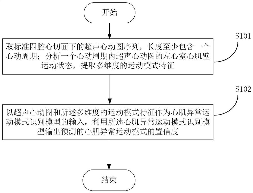
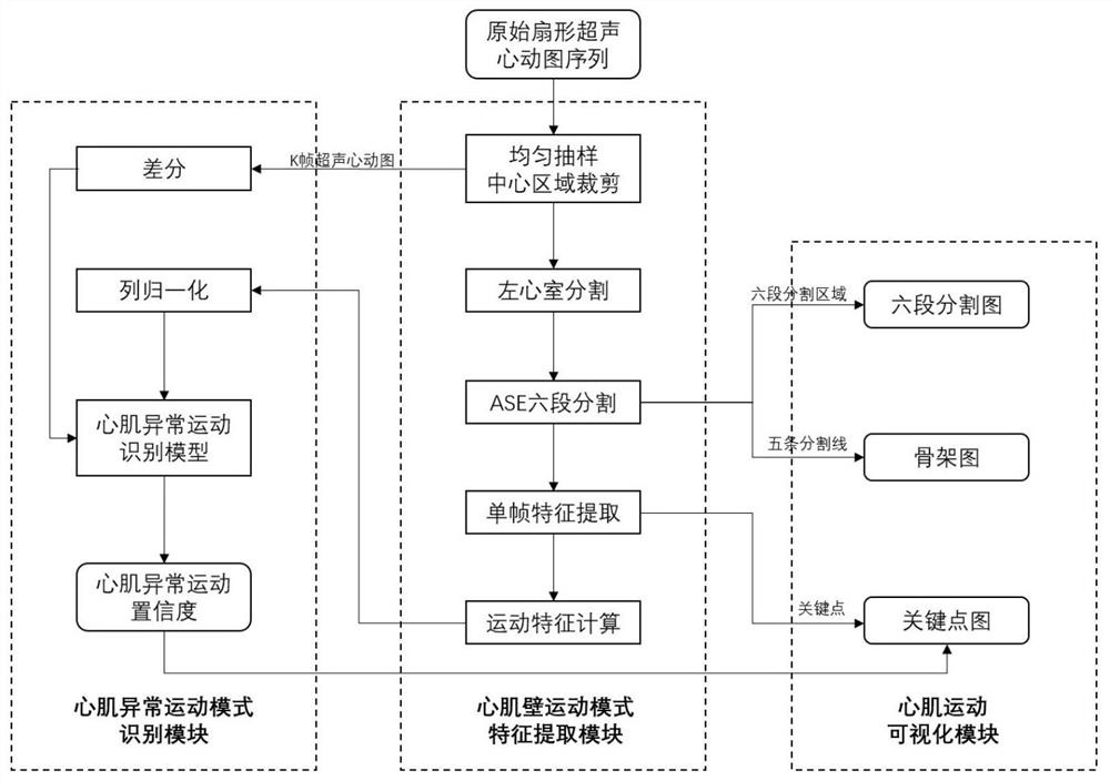
The invention provides an ultrasonic cardiogram myocardial abnormal motion mode analysis method and system and a storage medium and relates to the technical field of medical image processing. The method comprises the following steps of acquiring an echocardiogram sequence under a standard four-cavity cardiac tangent plane, wherein the length at least comprises one cardiac cycle; analyzing the leftventricular myocardial wall motion state of the echocardiogram in one cardiac cycle, and extracting multi-dimensional motion mode characteristics; and taking the echocardiogram sequence and the motion mode characteristics as the input of a myocardial abnormal motion mode identification model, and outputting the predicted confidence coefficient of the myocardial abnormal motion mode by using the myocardial abnormal motion mode identification model. According to the method, by extracting the motion mode characteristics in the echocardiogram sequence in one cardiac cycle, the time sequence information and the local motion information in the echocardiogram are fully mined, so accurate myocardial abnormal motion mode analysis can be automatically carried out according to the characteristics, and the prediction result is returned.

Description

technical field [0001] The invention relates to the technical field of medical image processing, in particular to a method, system and storage medium for analyzing abnormal myocardial motion patterns in echocardiography. Background technique [0002] An echocardiogram is a series of site-specific ultrasound images of the heart acquired using a phased array probe; it can noninvasively visualize the structure and motion patterns of the heart in real time. [0003] The analysis of echocardiograms is mainly done manually by sonographers or clinicians, which leads to an increasing workload for doctors. In addition, the accuracy of manual echocardiographic analysis largely depends on the doctor's experience, resulting in a certain degree of subjectivity in the analysis results. In recent years, with the rapid development of computer-related technologies and the maturity of graphics and image technology, the role and influence of medical image processing technology on medical rese...

Claims


Application Information

Patent Timeline
IPC IPC(8): G06T7/00G06T7/246G06T7/12G06T7/136G06T7/62G06T7/90
CPCG06T7/0016G06T2207/10132G06T2207/20081G06T2207/20084G06T2207/30048G06T7/12G06T7/136G06T7/246G06T7/62G06T7/90
Inventor 马壮张宾李雪包一平郭宝珠李宁于丹熊晟博
Owner 东软教育科技集团有限公司
Why Eureka
Try Eureka