White-matter high-signal grading method, electronic equipment and storage medium
A grading method and signal technology, applied in the field of image processing, can solve problems such as difficult to evaluate accurately, lack of brain partition function, etc., and achieve the effect of improving accuracy and efficiency
- Summary
- Abstract
- Description
- Claims
- Application Information
AI Technical Summary
Problems solved by technology
Method used
Image

Examples
Embodiment 1
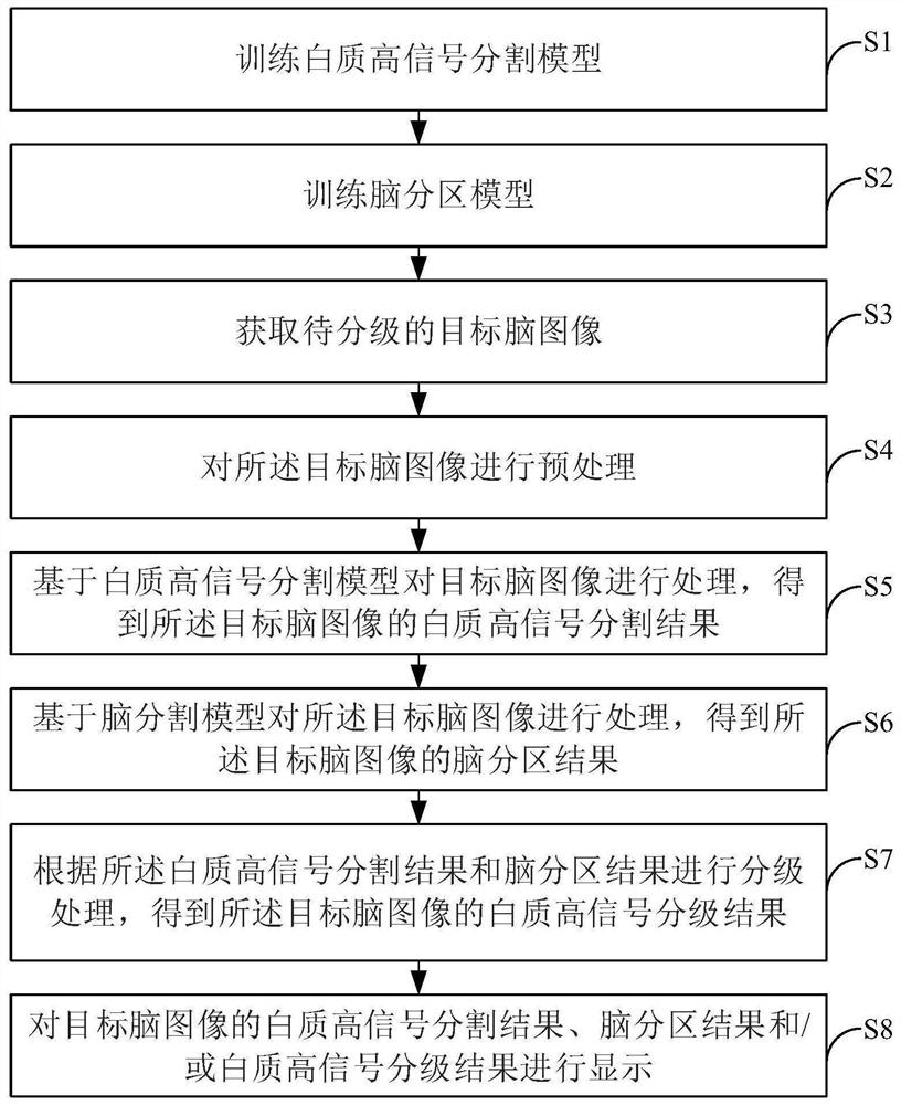
[0050] This embodiment provides a white matter hyperintensity classification method, such as figure 1 As shown, the method specifically includes the following steps:
[0051] S1, training a preset white matter hyperintensity segmentation model, which is used to obtain the white matter hyperintensity segmentation results of the target brain image.
[0052] In this embodiment, the white matter hyperintensity segmentation model is trained through the following steps:
[0053] S11. Acquire a first sample data set, where the first sample data set includes several first sample images and white matter hyperintensity tag labeling results corresponding to the first sample images. Specifically, the process of obtaining the first sample data set is as follows:
[0054] S111, acquire a first sample image, the first sample image may include but not limited to CT (Computed Tomography) image and MRI (Magnetic Resonance Imaging) image of the patient's head, where the MRI image may include F...
Embodiment 2
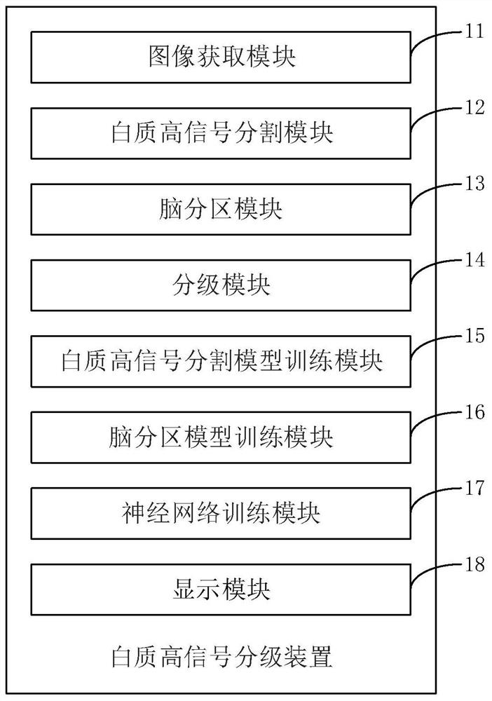
[0118] In this embodiment, the aforementioned step S7 is implemented as follows: use the pre-trained neural network to process the white matter hyperintensity segmentation results and brain partition results of the target brain image to obtain the white matter hyperintensity classification results of the target brain image.
[0119] As an example but not a limitation, the neural network of this embodiment can adopt a network structure such as VggNet, GoogleNet, ResNet, etc., whose input is the white matter hyperintensity segmentation result and brain partition result of the target brain image, and the output is the white matter hyperintensity classification prediction result.
[0120] In this embodiment, the neural network can be constructed and trained according to the scoring scale, and the training process of the neural network is as follows:
[0121] S71'. Acquire a fourth sample data set, where the fourth sample data set includes several fourth sample images and white mat...
Embodiment 3
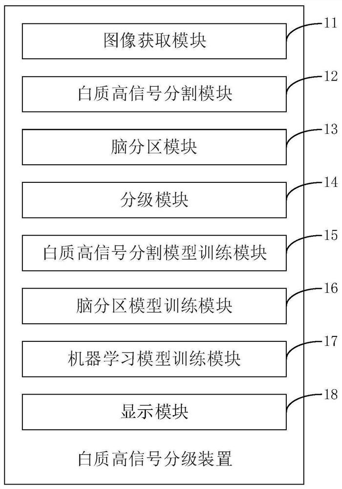
[0131] This embodiment provides a white matter hyperintensity grading device, such as figure 2 As shown, the device 1 specifically includes:
[0132] An image acquisition module 11, configured to acquire a target brain image;
[0133] A white matter hyperintensity segmentation module 12, configured to process the target brain image based on a pre-trained white matter hyperintensity segmentation model, to obtain a white matter hyperintensity segmentation result of the target brain image;
[0134] The brain partition module 13 is used to process the target brain image based on the pre-trained brain partition model to obtain the brain partition result of the target brain image;
[0135] The grading module 14 is configured to obtain a white matter hyperintensity classification result of the target brain image according to the white matter hyperintensity segmentation result and the brain partition result.
[0136] In this embodiment, the target brain image includes several slice...
PUM
 Abstract
Description
Claims
Application Information
 - R&D Engineer
- R&D Manager
- IP Professional
- Industry Leading Data Capabilities
- Powerful AI technology
- Patent DNA Extraction
Browse by: Latest US Patents, China's latest patents, Technical Efficacy Thesaurus, Application Domain, Technology Topic.
© 2024 PatSnap. All rights reserved.Legal|Privacy policy|Modern Slavery Act Transparency Statement|Sitemap