
Three-dimensional printing method of intracranial tumor, three-dimensional printing device and readable storage medium

A 3D printing technology for intracranial tumors, applied in image data processing, medical science, instruments, etc., can solve problems such as blurred 3D models, low penetration rate, and poor reflection of diseased tissues by 3D solid models, so as to improve Surgical success rate, ease of diagnosis and analysis

Active Publication Date: 2021-03-19
江门市中心医院
View PDF10 Cites 1 Cited by
  • Summary
  • Abstract
  • Description
  • Claims
  • Application Information

AI Technical Summary

Problems solved by technology

[0003] However, at present, the 3D models directly constructed from the scanned 2D images are relatively blurred, and the printed 3D solid models cannot reflect the diseased tissue well. For those operations that require high precision, such as intracranial tumor removal surgery, these The printed 3D solid model is not easy to use, so the penetration rate is not high

Method used


Image

  • Three-dimensional printing method of intracranial tumor, three-dimensional printing device and readable storage medium
  • Three-dimensional printing method of intracranial tumor, three-dimensional printing device and readable storage medium
  • Three-dimensional printing method of intracranial tumor, three-dimensional printing device and readable storage medium

Examples


Embodiment Construction

[0046] In order to make the objectives, technical solutions and advantages of the present invention, the present invention will be described in further detail below with reference to the accompanying drawings and examples. It will be appreciated that the specific embodiments described herein are intended to explain the present invention and is not intended to limit the invention.

[0047] It should be noted that although a functional module division is performed in the apparatus, a logical order is shown in the flowchart, but in some cases, it can be divided into modules different from the device, or in the order in the flowchart. The steps shown or described. The term "first", "second", or the like in the above appended claims are used to distinguish between similar objects without having to describe a particular order or ahead order.

[0048] The present invention provides a three-dimensional printing method, a three-dimensional printing apparatus, and a readable storage medium,...


PUM


Abstract

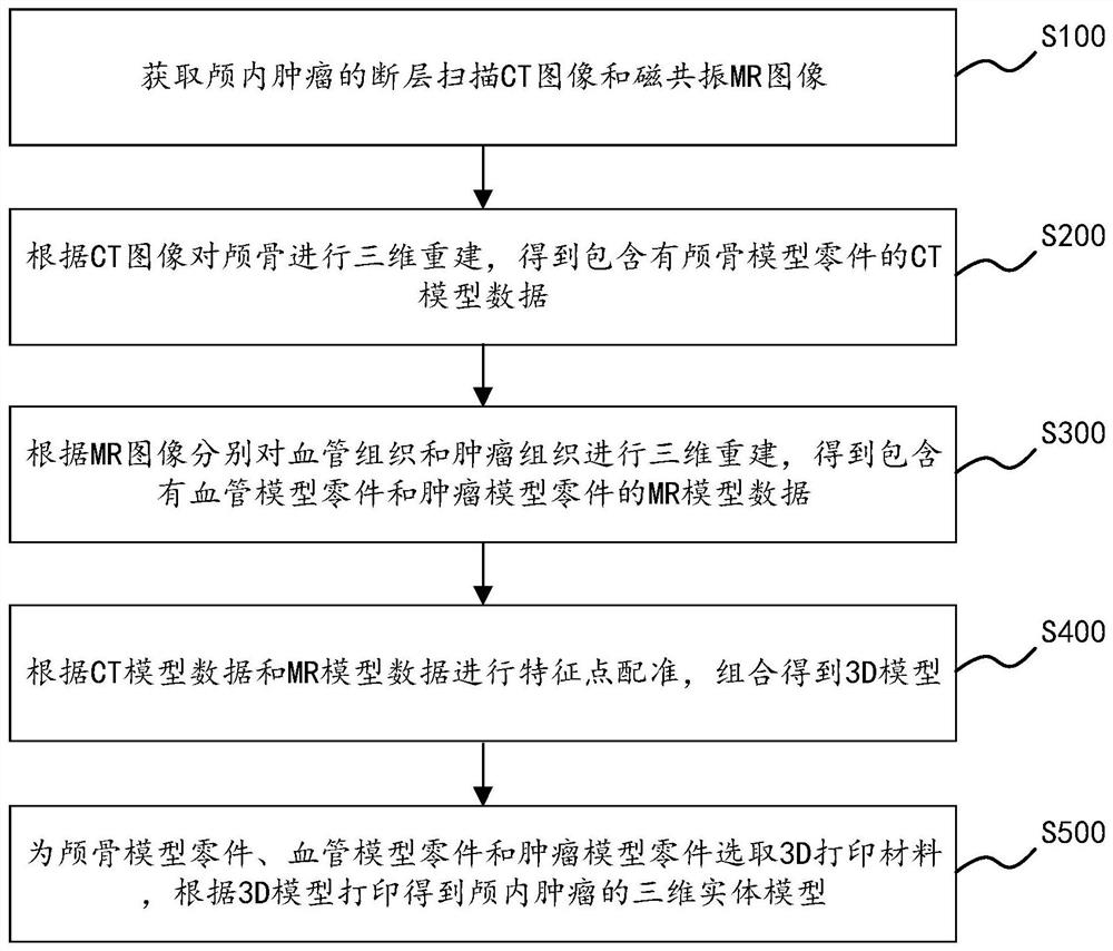
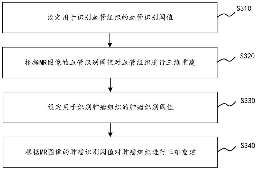
The invention discloses a three-dimensional printing method of intracranial tumor, a three-dimensional printing device and a readable storage medium. The three-dimensional printing method comprises the following steps: obtaining a computed tomography (CT) image and a magnetic resonance (MR) image of the intracranial tumor; performing three-dimensional reconstruction on a skull according to the CTimage to obtain CT model data; respectively carrying out three-dimensional reconstruction on vascular tissue and tumor tissue according to the MR image to obtain MR model data; performing feature point registration according to the CT model data and the MR model data, and performing combination to obtain a 3D model; and selecting a 3D printing material, and carrying out printing according to the 3D model to obtain a three-dimensional solid model of the intracranial tumor. 3D model construction is carried out on the skull, the vascular tissue and the tumor tissue by using respective characteristics of the CT image and the MR image, and the 3D model with high precision can be obtained, so that the 3D model printed by a 3D printer can accurately reflect the situation of the tumor tissue in the skull, and a doctor can diagnose and analyze a patient conveniently.

Description

Technical field [0001] The present invention relates to the field of three-dimensional printing techniques, and more particularly to a three-dimensional printing method, a three-dimensional printing device, and a readable storage medium for intracranial tumors. Background technique [0002] 3D printing, also known as 3D printing, a three-dimensional printing technology in recent years; with the increasing popularity of three-dimensional printing technology, more and more items can be printed through 3D printing technology. Among them, in the field of clinical medicine, the two-dimensional scan image of the human tissue constitutes a 3D model, and the human tissue can be printed by means of 3D printing techniques to facilitate the diagnosis and analysis of different tissues for different patients. [0003] However, the three-dimensional model directly constructed by scanned two-dimensional image is relatively blurred, and the 3D solid model of printing does not reflect the lesion,...

Claims


Application Information

Patent Timeline
IPC IPC(8): A61B34/10B33Y30/00B33Y50/00B33Y80/00G06T7/33G06T17/00B33Y70/00
CPCG06T17/00G06T7/33A61B34/10B33Y30/00B33Y50/00B33Y80/00B33Y70/00G06T2207/10081G06T2207/10088G06T2207/30096A61B2034/105
Inventor 董家军伍益彭逸龙
Owner 江门市中心医院
Learn More