Tumor prediction method of PET-CT image based on neural network and computer readable storage medium

A PET-CT, CT image technology, applied in the field of PET-CT equipment, can solve the problem of not being able to make good use of PET and CT, and achieve the effects of improving recognition accuracy, increasing accuracy and reducing work efficiency

Pending Publication Date: 2021-04-20
浙江明峰智能医疗科技有限公司
View PDF3 Cites 3 Cited by
  • Summary
  • Abstract
  • Description
  • Claims
  • Application Information

AI Technical Summary

Problems solved by technology

[0004] The current tumor segmentation task is mainly based on the Unet network. The input data is basically to splice PET and CT into dual-channel image data for training. However, this network cannot make good use of the information of PET and CT.

Method used


Image

  • Tumor prediction method of PET-CT image based on neural network and computer readable storage medium
  • Tumor prediction method of PET-CT image based on neural network and computer readable storage medium
  • Tumor prediction method of PET-CT image based on neural network and computer readable storage medium

Examples


Embodiment Construction

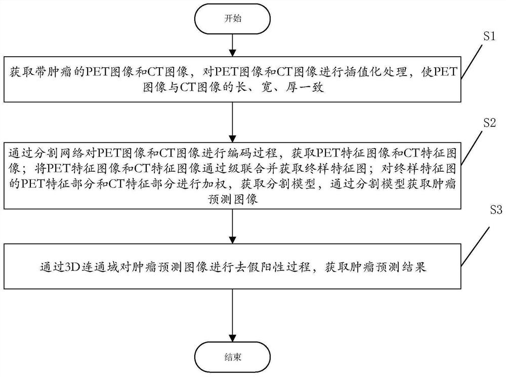
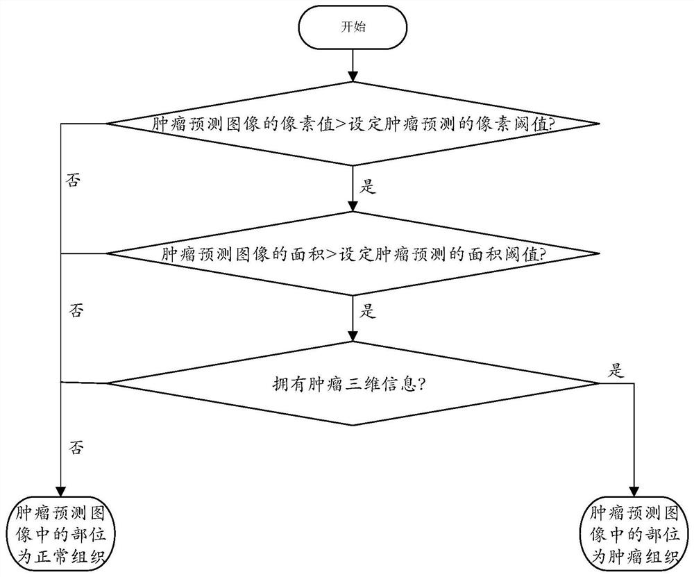
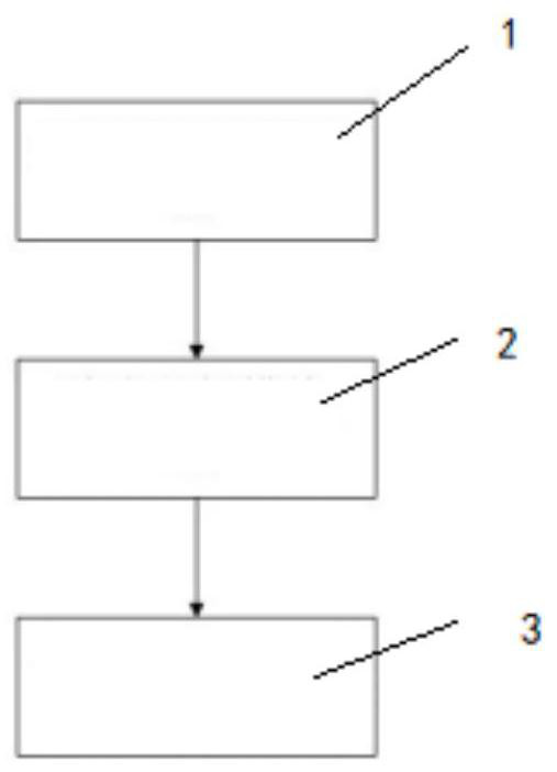
[0027]The advantages of the present invention will be further illustrated below with reference to the accompanying drawings.

[0028]The exemplary embodiment will be described in detail herein, and examples thereof are shown in the drawings. The following description is related to the drawings, unless otherwise indicated, the same numbers in the drawings represent the same or similar elements. The embodiments described in the exemplary embodiments described below do not represent all embodiments consistent with the present disclosure. Instead, they are only examples of devices and methods consistent with some aspects of the present disclosure as detailed in the appended claims.

[0029]The terms used in the present disclosure are only for the purpose of describing particular embodiments, not intended to limit the disclosure. "One", "one", "one", "one", "one", "" "and" "" as used in the present disclosure and the appended claims are also intended to include other forms unless the context c...


PUM


Abstract

The invention discloses a tumor prediction method for a PET-CT image based on a neural network. The tumor prediction method comprises the following steps: carrying out interpolation processing on a PET image and a CT image, wherein the lengths, widths and thicknesses of the two images are consistent; carrying out a coding process on the PET image and the CT image through a segmentation network, obtaining a PET feature image and a CT feature image, carrying out cascade combination, obtaining a final sample feature map, carrying out weighting on a PET feature part and a CT feature part of the final sample feature map, obtaining a segmentation model, and obtaining a tumor prediction image through the segmentation model; and performing a false positive removal process on the tumor prediction image through a 3D connected domain to obtain a tumor prediction result. The tumor is automatically segmented and identified by using the deep learning technology of the neural network, the suspected tumor tissue can be rapidly identified, the part is directly analyzed by a doctor, and finally, the diagnosis result is determined by the doctor, so that the working efficiency of a radiologist can be greatly reduced, and the tumor identification accuracy is improved.

Description

Technical field[0001]The present invention relates to the technical field of PET-CT equipment, and more particularly to a tumor prediction method and a computer readable storage medium based on a neural network-CT image.Background technique[0002]The PET-CT scanner is a high-end nuclear medicine imaging device that combines PET and CT. CT mainly uses different tissue in the human body to imaging different absorption strength of X-rays; PET mainly transmits positron radioactivity, can track the abnormal glucose metabolism process in vivo. PET-CT can better display the position and size information of the tumor with CT alone.[0003]Due to the lack of a large number of radiologists in all hospitals in my country, a radiologist needs to look at hundreds of radioblasts every day, bringing a huge work burden to the radiologist, and the intensity of full load can also increase the doctor's tumor. The misdiagnosis rate. And the diagnosis of different working experiences may have a distinct re...

Claims


Application Information

Patent Timeline
Patent Type & Authority Applications(China)
IPC IPC(8): G06T7/00G06T7/10G06N3/04G06N3/08
Inventor 赵东东王小状叶宏伟
Owner 浙江明峰智能医疗科技有限公司
Why Eureka
Try Eureka