Method and equipment for measuring bone mineral density through oral cavity cone beam CT
A cone beam, bone density technology, used in radiological diagnostic instruments, medical science, dental radiological diagnosis, etc., can solve problems such as the inability to directly obtain the density image of bone tissue, affecting the success rate of surgery, and errors.
- Summary
- Abstract
- Description
- Claims
- Application Information
AI Technical Summary
Problems solved by technology
Method used
Image
Examples
Embodiment 1
[0088] A method for measuring bone density by oral cone beam CT, comprising the steps of:
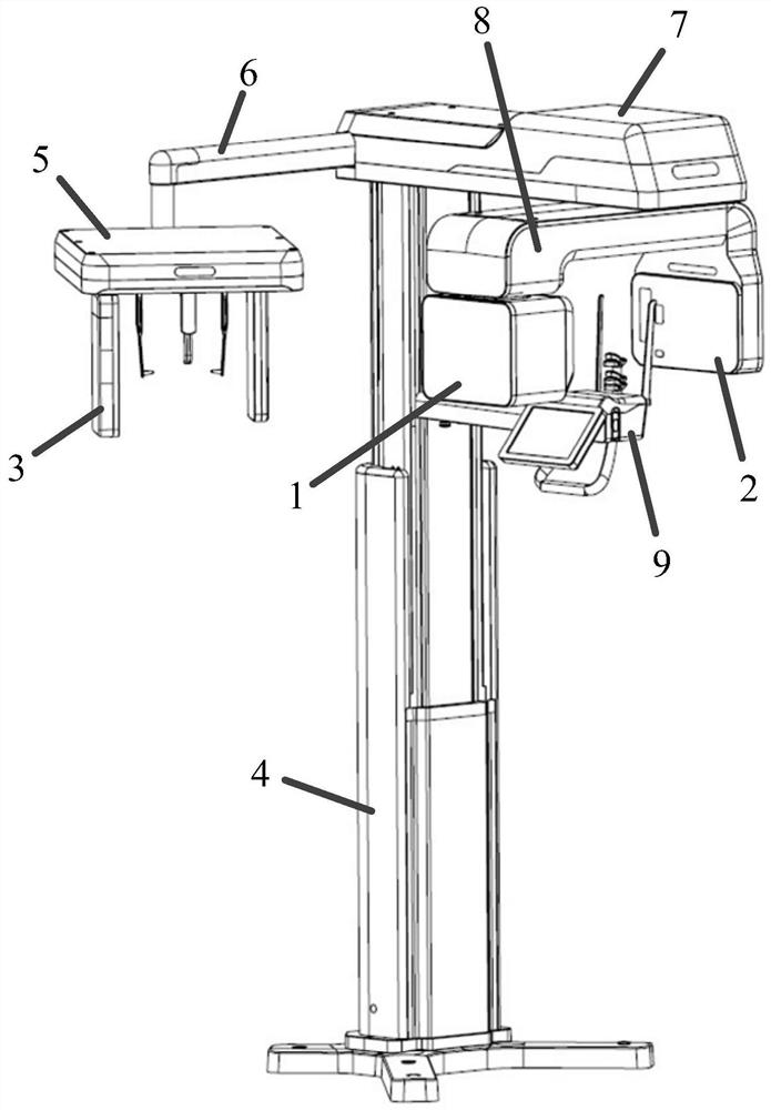
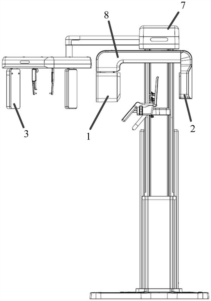
[0089] S1, sending X-rays to the scanned body from different angles through the oral cone beam CT equipment, so as to obtain the projection information of the scanned body at different angles;
[0090] S2. Obtain high-energy X-rays and low-energy X-rays passing through the scanned body, respectively;
[0091] S3. The processing unit separately acquires high-energy X-ray signals and low-energy X-ray signals passing through the scanned body, and uses an image reconstruction algorithm to obtain a bone tissue density image.
[0092] In this embodiment, the method for measuring oral bone density, in step S3, also includes:
[0093] According to the reconstruction results of the projection data of the high-energy X-ray and the low-energy X-ray under the high-voltage exposure condition and the low-voltage exposure condition respectively, the decomposition coefficient C of bone tissue is respe...
Embodiment 2
[0110] A method for measuring bone density by oral cone beam CT, comprising the steps of:
[0111] S1, sending X-rays to the scanned body from different angles through the oral cone beam CT device, thereby obtaining the projection information of the scanned body at different angles; in step S1, including: two ray sources; one of the ray sources Under high-voltage exposure conditions, high-energy X-rays are sent to teeth to obtain a set of high-energy attenuation data; another ray source sends low-energy X-rays to teeth under low-voltage exposure conditions to obtain a set of low-energy attenuation data;
[0112] S2. Obtain high-energy X-rays and low-energy X-rays passing through the scanned body, respectively;
[0113] S3. The processing unit separately acquires high-energy X-rays and low-energy X-rays passing through the scanned body, and uses an image reconstruction algorithm to obtain a bone tissue density image.
Embodiment 3
[0115] A method for measuring bone density by oral cone beam CT, comprising the steps of:
[0116] S1, sending X-rays to the scanned body from different angles through the oral cone beam CT device, thereby obtaining the projection information of the scanned body at different angles; in step S1, including: a ray source; during one scanning process, Send X-rays to the scanned object under high-voltage exposure conditions to obtain a set of high-energy attenuation data; in another scanning process, send X-rays to the scanned object under low-voltage exposure conditions to obtain a set of low-energy attenuation data;
[0117] S2. Obtain high-energy X-rays and low-energy X-rays passing through the scanned body, respectively;
[0118] S3. The processing unit separately acquires high-energy X-rays and low-energy X-rays passing through the scanned body, and uses an image reconstruction algorithm to obtain a bone tissue density image.
PUM
 Abstract
Description
Claims
Application Information
 - R&D
- Intellectual Property
- Life Sciences
- Materials
- Tech Scout
- Unparalleled Data Quality
- Higher Quality Content
- 60% Fewer Hallucinations
Browse by: Latest US Patents, China's latest patents, Technical Efficacy Thesaurus, Application Domain, Technology Topic, Popular Technical Reports.
© 2025 PatSnap. All rights reserved.Legal|Privacy policy|Modern Slavery Act Transparency Statement|Sitemap|About US| Contact US: help@patsnap.com


