Anti-PD-L1/CD47 bispecific antibody and use thereof

A bispecific antibody, PD-L1 technology, used in the field of biomedicine or biopharmaceuticals

Active Publication Date: 2021-05-04
SHANGHAI NOVAMAB BIOPHARM CO LTD
View PDF7 Cites 4 Cited by
  • Summary
  • Abstract
  • Description
  • Claims
  • Application Information

AI Technical Summary

Problems solved by technology

[0005] So far, there is no bispecific single-domain antibody targeting PD-L1/CD47 on the market. As a new force in the diagnosis and treatment of a new generation of antibodies, single-domain antibodies have high s...

Method used


Image

  • Anti-PD-L1/CD47 bispecific antibody and use thereof
  • Anti-PD-L1/CD47 bispecific antibody and use thereof
  • Anti-PD-L1/CD47 bispecific antibody and use thereof

Examples


Embodiment 1

[0199] Example 1: Molecular structure design and expression of PD-L1 / CD47 bispecific antibody

[0200] The inventors designed bispecific antibodies that simultaneously bind human PD-L1 and CD47 extracellular domains. Among them, the PD-L1 single domain antibody sequence is derived from Chinese patent CN201810151835.5, and the CD47 single domain antibody sequence is derived from Chinese patent CN201810151752.6.

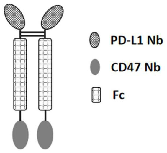
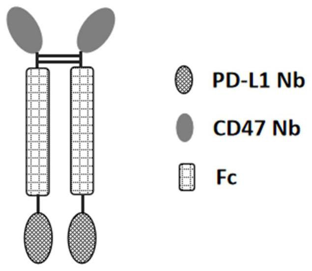
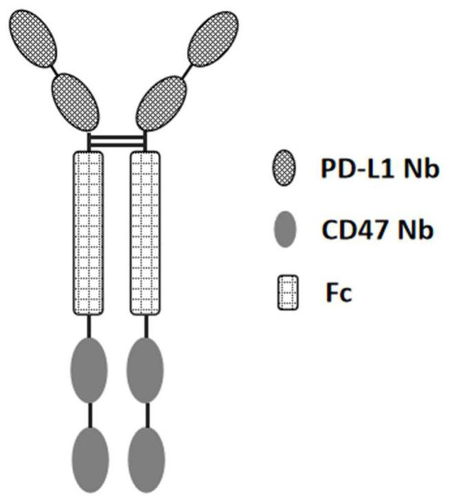
[0201] The present invention designs a variety of PD-L1 / CD47 bispecific antibody molecules with different structures, and measures their activity. This example only exemplifies the bispecific antibody molecules with four structures, which are as follows: Figure 1A The indicated double antibody A, such as Figure 1B The double antibody B shown, such as Figure 1C Double Antibody C as indicated and as Figure 1D In the shown double antibody D, double antibody A and double antibody B are in the form of tetravalent antibodies, and double antibody C and double antibody D...

Embodiment 2

[0205] Example 2: Comparison of blocking activity of PD-L1 / CD47 bispecific antibody on CD47 / SIRPa pathway

[0206] (1) Each sample takes 5×10 5 In 0.5% BSA-PBS buffer, several Jurkat cells (highly expressing human CD47) were added with serially diluted PD-L1 / CD47 bispecific antibody and positive control CD47 single domain antibody Fc fusion protein (Nb-Fc), antibody dilution gradient For 514nM, 257nM, 129nM, 64.3nM, 32.1nM, 16.1nM, 8.03nM, 4.02nM, 2.00nM, 1.00nM, 0.50nM, 0.25nM, add 100uL to each sample, and add 5ug hSIRPa(ECD) to all samples at the same time -Fc-Biotin, incubated at 4°C for 20 min; (2) Wash the cells twice with PBS, add eBioscience SA-PE, incubate at 4°C for 20 min, wash the cells twice with PBS, and detect them with a flow cytometer (BD FACS Calibur). Graphpad Prism 6 software was used for data processing.

[0207] The result is as figure 2 Shown: The blocking activity of the octavalent antibody is better than that of the tetravalent antibody, and the te...

Embodiment 3

[0208] Example 3: Comparison of blocking activity of PD-L1 / CD47 bispecific antibody on PD-1 / PD-L1 pathway

[0209] Take 3×10 for each sample 5 A375 / PD-L1 stably transfected cells were added to 0.5% BSA-PBS buffer, and the double antibody to be tested and the control antibody PD-L1 single domain antibody Fc fusion protein (Nb-Fc) were added in a serial dilution, and the antibody dilution gradient was 184nM, 91.8nM, 45.9nM, 22.9nM, 11.5nM, 5.74nM, 2.87nM, 1.43nM, 0.72nM, 0.36nM, 0.18nM, 0.09nM, 100uL was added to each sample, and 3ug hPD-1(ECD )-Fc-Biotin, incubated at 4°C for 20 min; (2) Wash the cells twice with PBS, add the SA-PE of eBioscience, incubate at 4°C for 20 min, wash the cells twice with PBS, and detect them with a flow cytometer (BDFACS Calibur). Graphpad Prism 6 software was used for data processing.

[0210] The result is as image 3 As shown: the bispecific antibodies with different structures have lower PD1 / PD-L1 interaction blocking activity than the origi...


PUM


Abstract

The invention provides an anti-PD-L1/CD47 bispecific antibody and use thereof. Specifically, the invention provides a bispecific antibody which comprises (a) a PD-L1 single-domain antibody and (b) a CD47 single-domain antibody. The invention provides a coding sequence for coding the bispecific antibody, a corresponding expression vector, a host cell capable of expressing the bispecific antibody, and a production method of the bispecific antibody. The bispecific antibody can target PD-L1 and CD47 at the same time, can not only activate T cells, but also effectively promote phagocytosis of Jurkat cells by macrophages without causing human red blood cell agglutination, and has good application prospects.

Description

technical field [0001] The present invention relates to the technical field of biomedicine or biopharmaceuticals, and more specifically relates to an anti-PD-L1 / CD47 bispecific antibody and its application. Background technique [0002] The body's tumor immune response is the result of the combined effect of the host's immune function status and tumor immunogenicity. Tumor cells can cause immune escape of tumor cells through abnormal expression of immunosuppressive molecules. Recent studies have shown that the PD-1 / PD-L1 inhibitory pathway plays an important role in tumor immune response, mediation of transplant rejection, occurrence and development of autoimmune diseases, and chronic viral infection. PD-L1 is expressed on the surface of activated T cells, B cells, macrophages, dendritic cells, tumor cells, etc., or immune shielding tissues (placenta, vascular endothelium, etc.). Antibodies targeting PD-1 / PD-L1 promote T cell proliferation and cytokine release in vitro by ...

Claims


Application Information

Patent Timeline
IPC IPC(8): C07K16/46C12N15/13G01N33/68G01N33/574A61K47/68A61P35/00
CPCC07K16/2827C07K16/2803A61K47/6849A61P35/00G01N33/6872G01N33/57492C07K2319/30C07K2319/33C07K2317/31C07K2317/73C07K2317/92C07K2317/94
Inventor 万亚坤朱敏盖军伟沈晓宁
Owner SHANGHAI NOVAMAB BIOPHARM CO LTD
Why Eureka
Try Eureka