
Trachea cannula fixator for first aid in intensive care medicine department

A technology of endotracheal intubation and fixer, which is applied in the field of medical devices, can solve the problems of patients' teeth injury, epidermis ulceration, difficult replacement, etc., and achieve the effect of reducing contact area, reducing oppression, and convenient fixation

Inactive Publication Date: 2021-07-13
SECOND AFFILIATED HOSPITAL OF COLLEGE OF MEDICINEOF XIAN JIAOTONG UNIV
View PDF0 Cites 1 Cited by
  • Summary
  • Abstract
  • Description
  • Claims
  • Application Information

AI Technical Summary

Problems solved by technology

[0002] Endotracheal intubation is one of the important measures for successfully rescuing critically ill patients. The traditional fixation method for endotracheal intubation is to fix the dental pad and endotracheal tube with adhesive plaster and then paste them directly to the cheeks on both sides of the patient. Due to the crude fixation method, Therefore, there are many disadvantages. Fixing with adhesive plaster often makes the patient feel uncomfortable, stimulates the skin and causes inflammation or epidermal ulceration. If the adhesive plaster is left for too long, it is easy to stick together and form a group, which is not easy to replace, and it is very easy to pollute and cause secondary infection to occur, causing serious harm to the patient. Extra pain, in order to prevent the patient from biting and closing the trachea, the existing tracheal tube holders are all made of hard materials. Although this avoids the problem of occlusion, it is easy to cause damage to the patient's teeth, so a serious need is urgently needed. Endotracheal Tube Holder

Method used


Image

  • Trachea cannula fixator for first aid in intensive care medicine department
  • Trachea cannula fixator for first aid in intensive care medicine department
  • Trachea cannula fixator for first aid in intensive care medicine department

Examples


Embodiment Construction

[0025] The present invention will be further described below in conjunction with the accompanying drawings.

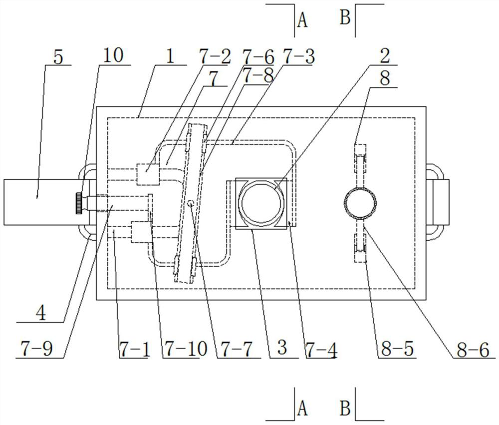
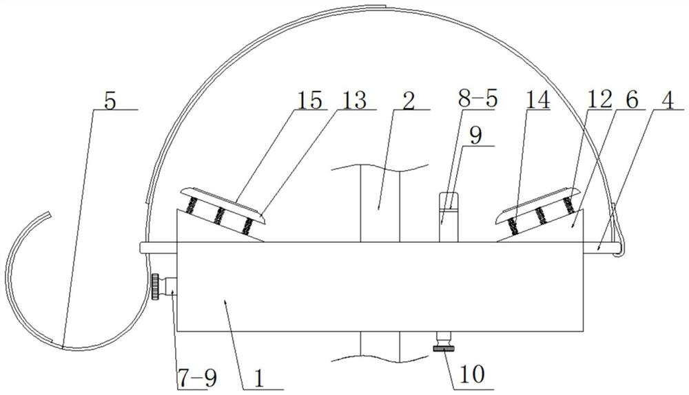
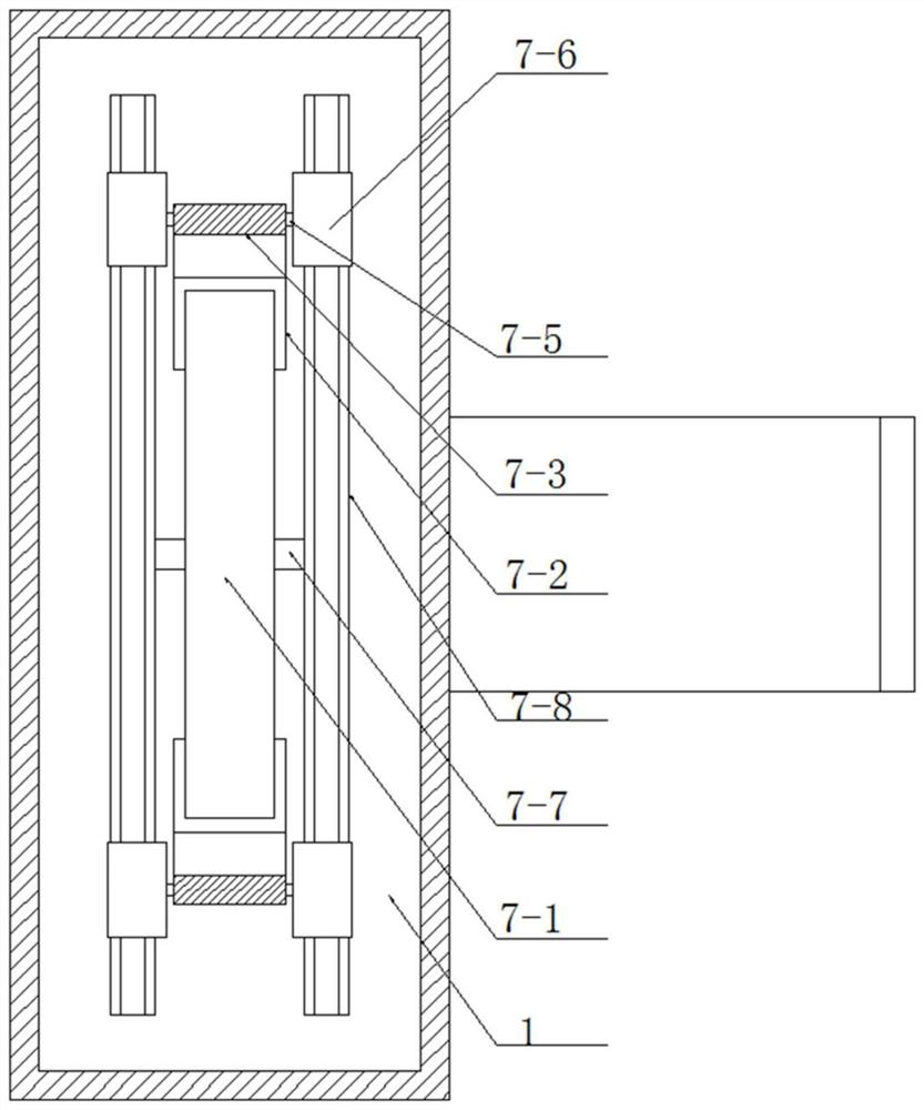
[0026] see as Figure 1-Figure 5 As shown, the technical solution adopted in this specific embodiment is: it includes a box body 1 and an endotracheal tube 2, through holes 3 are symmetrically opened on the front and rear side walls of the box body 1, and the endotracheal tube 2 is movably inserted in the box body 1 Inside the through hole 3 on the front and rear side walls; it also includes a fixing buckle 4, a strap 5, an endotracheal tube clamping mechanism 7 and an opening mechanism 8; the left and right side walls of the box body 1 are symmetrically connected with fixing buckles by bolts 4. One end of the strap 5 is fixedly bound to the fixed buckle 4 on the right, and the other end of the strap 5 moves through the fixed buckle 4 on the left, and passes through the strap 5 between the Velcro and the two fixed buckles 4 The outer side wall is fixedly connected, an...


PUM


Abstract

The invention provides a trachea cannula fixator for first aid in the intensive care medicine department, and relates to the technical field of medical instruments. The trachea cannula fixator comprises a box body and trachea cannulas, through holes are symmetrically formed in the front side wall and the rear side wall of the box body, and the trachea cannulas are movably inserted into the through holes in the front side wall and the rear side wall of the box body; the trachea cannula fixator further comprises a fixing buckle, a bandage, a trachea cannula clamping mechanism and a mouth opening mechanism. Fixing buckles are symmetrically and fixedly arranged on the left side wall and the right side wall of the box body, one end of the bandage is fixedly connected with the fixing buckle on the right side in a binding mode, and the other end of the bandage movably penetrates through the fixing buckle on the left side and then is fixedly connected with the outer side wall, between the two fixing buckles, of the bandage through hook-and-loop fasteners; triangular supporting blocks are symmetrically and fixedly arranged on the left side and the right side of the rear side wall of the box body. The trachea cannula clamping mechanism is arranged on the left side in the box body, the mouth opening mechanism is arranged on the right side in the box body, the trachea cannula can be stably fixed, operation is simple, meanwhile, the oral cavity of the patient can be opened, and the situation that the ventilation of the trachea cannula is affected due to tooth occlusion is prevented.

Description

technical field [0001] The invention relates to the technical field of medical devices, in particular to a tracheal intubation fixer for first aid in the intensive care department. Background technique [0002] Endotracheal intubation is one of the important measures for successfully rescuing critically ill patients. The traditional fixation method for endotracheal intubation is to fix the dental pad and endotracheal tube with adhesive plaster and then paste them directly to the cheeks on both sides of the patient. Due to the crude fixation method, Therefore, there are many disadvantages. Fixing with adhesive plaster often makes the patient feel uncomfortable, stimulates the skin and causes inflammation or epidermal ulceration. If the adhesive plaster is left for too long, it is easy to stick together and form a group, which is not easy to replace, and it is very easy to pollute and cause secondary infection to occur, causing serious harm to the patient. Extra pain, in order...

Claims


Application Information

Patent Timeline
IPC IPC(8): A61M16/04
CPCA61M16/0434A61M16/0493
Inventor 韩俊丽骆艳妮张慧藩赵潇
Owner SECOND AFFILIATED HOSPITAL OF COLLEGE OF MEDICINEOF XIAN JIAOTONG UNIV
Learn More