Image data-based segmentation method, device and equipment

A technology of image data and data, applied in the field of medical imaging and computer, can solve the problems of poor bone removal effect, increase the radiation dose of patients, distinguish calcification parts, etc., and achieve the effect of high accuracy and saving analysis time.

Active Publication Date: 2021-08-03
UNION STRONG (BEIJING) TECH CO LTD
View PDF10 Cites 1 Cited by
  • Summary
  • Abstract
  • Description
  • Claims
  • Application Information

AI Technical Summary

Problems solved by technology

[0005] The embodiment of this specification provides a segmentation method, device, and equipment based on image data, which are used to solve the following technical problems: two CT scans are required, which increases the radiation dose of the patient; the position of the bone removal image does not match easily, resulting in bone removal The bone effect becomes worse, and even needs to be re-scanned; the error is large, and it is difficult to distinguish the calcified part from the bone tissue

Method used


Image

  • Image data-based segmentation method, device and equipment
  • Image data-based segmentation method, device and equipment
  • Image data-based segmentation method, device and equipment

Examples


Embodiment Construction

[0087] In order to enable those skilled in the art to better understand the technical solutions in this specification, the technical solutions in the embodiments of this specification will be clearly and completely described below in conjunction with the drawings in the embodiments of this specification. Obviously, the described The embodiments are only some of the embodiments of the present application, but not all of them. Based on the embodiments of this specification, all other embodiments obtained by persons of ordinary skill in the art without creative efforts shall fall within the scope of protection of this application.

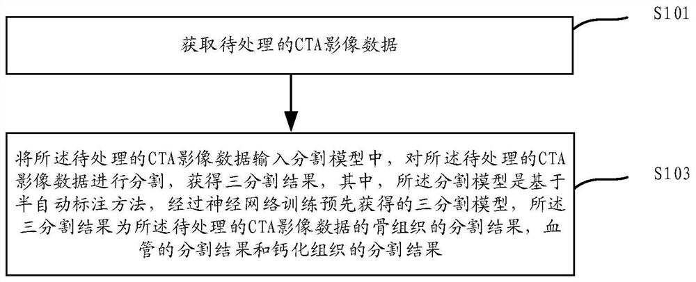
[0088] figure 1 A schematic diagram of a segmentation method based on image data provided by the embodiment of this specification, such as figure 1 As shown, the method includes:

[0089] Step S101: Obtain CTA image data to be processed.

[0090] This manual uses CTA image data as the object to be processed to realize the segmentation of bone tissu...


PUM


Abstract

The embodiment of the invention discloses a segmentation method, device and equipment based on image data. The method comprises the following steps that CTA image data to be processed is acquired; the to-be-processed CTA image data are input into a segmentation model, the to-be-processed CTA image data are segmented, a three-segmentation result is obtained, the segmentation model is a three-segmentation model obtained in advance through neural network training on the basis of a semi-automatic labeling method, the three segmentation results are a bone tissue segmentation result, a blood vessel segmentation result and a calcified tissue segmentation result of the to-be-processed CTA image data. By adopting the segmentation method provided by the specification, the segmentation of bone tissues, calcified tissues and blood vessels can be automatically realized, the analysis time is greatly saved, and the accuracy is relatively high.

Description

technical field [0001] This specification relates to the field of medical imaging and computer technology, in particular to a segmentation method, device and equipment based on image data. Background technique [0002] CTA (computed tomography angiography) image data has important clinical applications and can be used as an auxiliary diagnostic means for the clinical treatment of intracranial aneurysms. Since the bone tissue in brain imaging will affect the clinical diagnosis results, in order to achieve better diagnostic results and reduce diagnostic errors, it is often necessary to remove bone tissue in brain imaging data. [0003] In the prior art, for bone removal of CTA image data, two sets of images are generally used, and bone tissue is removed through differential processing. Specifically, using non-contrast CT image data (NCCT) and CTA image data, wherein, the NCCT image is an image that does not need to be injected with a contrast agent, and the CTA image is an im...

Claims


Application Information

Patent Timeline
Patent Type & Authority Applications(China)
IPC IPC(8): G06T7/00G06T7/11G06T7/136G06T7/70
CPCG06T7/0012G06T7/11G06T7/136G06T7/70G06T2207/10081G06T2207/30101G06T2207/30008G06T2211/404G06T2207/20081G06T2207/20084
Inventor 方刚宋凌印胤杨光明
Owner UNION STRONG (BEIJING) TECH CO LTD
Why Eureka
Try Eureka