Gram staining leucorrhea smear color microscopic image segmentation method and system

A Gram staining and microscopic image technology, which is applied in image analysis, image enhancement, image data processing, etc., can solve the problems of separation of negative and positive targets, adhesion and mixing of negative and positive targets, and achieve the effect of ensuring accuracy

Pending Publication Date: 2021-08-03
JIANGSU BIOPERFECTUS TECH CO LTD
View PDF3 Cites 1 Cited by
  • Summary
  • Abstract
  • Description
  • Claims
  • Application Information

AI Technical Summary

Problems solved by technology

[0004] In the prior art, since the Gram-stained high-magnification leucorrhea dry film color microscopic image contains a large amount of information, including independent target cells, adherent target cells, impurities and other unknown substances, it is necessary to detect the target before detecting the target. Image preprocessing is performed on the image. In the process of processing, there are large areas of negative and positive targets sticking together and mixed together. It is difficult to accurately separate the negative and positive targets using traditional target segmentation methods. In order to obtain accurate morphological features of the target, Targeted image segmentation is required for the fixed color features of negative and positive targets

Method used


Image

  • Gram staining leucorrhea smear color microscopic image segmentation method and system
  • Gram staining leucorrhea smear color microscopic image segmentation method and system
  • Gram staining leucorrhea smear color microscopic image segmentation method and system

Examples


Embodiment Construction

[0059] The present invention will be described in detail below in conjunction with specific embodiments. The following examples will help those skilled in the art to further understand the present invention, but do not limit the present invention in any form. It should be noted that those skilled in the art can make several changes and improvements without departing from the concept of the present invention. These all belong to the protection scope of the present invention.

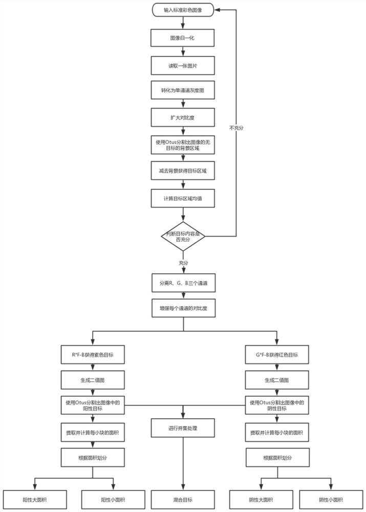
[0060] The embodiment of the present invention provides a kind of Gram-stained leucorrhea smear color microscopic image segmentation method, referring to figure 1 As shown, the specific steps are as follows:

[0061] First of all, collect and input the Gram-stained leucorrhea standard smear, which can be collected with a 100 times oil lens and a high-definition color camera in this embodiment. After the required cell image is collected, the method of downsampling the collected cell image is as follows:...


PUM


Abstract

The invention provides a Gram staining leucorrhea smear color microscopic image segmentation method and system, and relates to the technical field of digital image processing, and the method comprises the steps: collecting and inputting a Gram staining leucorrhea standard smear; carrying out downsampling and normalization on the collected cell image to obtain a standard size image; converting the standard size image into a single-channel grayscale image; processing the single-channel grayscale image, and excluding a target-free image; separating R, G and B channels in the color image, and performing normalization processing; obtaining and calculating an intersection part of the positive target area and the negative target area, and taking a union set as a mixed negative and positive target; cutting an area in which the negative and positive targets are mixed to form all independent negative and positive targets; and obtaining and classifying area information of each independent target, carrying out targeted form recognition, and counting the number to output a recognition result. According to the invention, possibility can be provided for subsequent cellular morphology recognition, and the preprocessing problem of an input image in target recognition is solved.

Description

technical field [0001] The invention relates to the technical field of digital image processing, in particular to a method and system for segmenting color microscopic images of Gram-stained leucorrhea smears. Background technique [0002] Leucorrhea is the secretion of female vagina, and the detection of white blood cells in leucorrhea is an important condition for judging whether the female reproductive system is healthy. Most of the traditional microscopic cell detection is manual microscopic examination. This method takes a long time to operate, has high work intensity, is easy to pollute the environment, and is subject to subjective influence, which is not conducive to clinical diagnosis. In recent years, with the development of artificial intelligence research and the improvement of biomedical image processing technology, it has become a reality to use image processing technology to automatically identify the formed components of microscopic cell images. In the face of...

Claims


Application Information

Patent Timeline
Patent Type & Authority Applications(China)
IPC IPC(8): G06T7/11G06T7/136G06T7/62G06T7/13G06T7/194G06T7/90
CPCG06T7/11G06T7/136G06T7/62G06T7/90G06T7/194G06T7/13G06T2207/10061G06T2207/30024G06T2207/30242
Inventor 娄博华朱慧敏朱子豪沈海东刘中华王国强
Owner JIANGSU BIOPERFECTUS TECH CO LTD
Why Eureka
Try Eureka