Devices, systems, and methods for tumor visualization and removal

A technology of imaging device and structure, applied in the direction of preparations, applications, pharmaceutical formulations, etc. for in vivo experiments, can solve few problems such as intraoperative histopathology

Pending Publication Date: 2021-11-05
SBI 艾拉制药加拿大有限公司
View PDF6 Cites 0 Cited by
  • Summary
  • Abstract
  • Description
  • Claims
  • Application Information

AI Technical Summary

Problems solved by technology

[0007] Current best practice for BCS involves palpation and / or specimen radiography with little intraoperative histopathology to guide removal

Method used


Image

  • Devices, systems, and methods for tumor visualization and removal
  • Devices, systems, and methods for tumor visualization and removal
  • Devices, systems, and methods for tumor visualization and removal

Examples


Embodiment Construction

[0039]Existing surgical margin assessment techniques focus on ex vivo samples to determine whether the surgical margin includes residual cancer cells. These techniques are limited by their inability to accurately spatially co-localize positive edges detected on ex vivo samples to the surgical bed, and the present disclosure overcomes this limitation by directly imaging the surgical cavity.

[0040] Other non-targeted techniques for reducing re-removal include studies combining non-targeted margins with standard-of-care BCS. While this technique may reduce the overall number of re-removals, the method includes several potential disadvantages. For example, larger resections are associated with poorer cosmetic outcomes, and non-targeted removal of extra tissue contradicts the intent of BCS. Furthermore, the end result of using such a technique appears to be in conflict with the recently updated ASTRO / SSO guidelines, which define a positive margin as "tumor at the ink" and do not...


PUM


Abstract

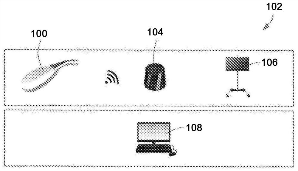
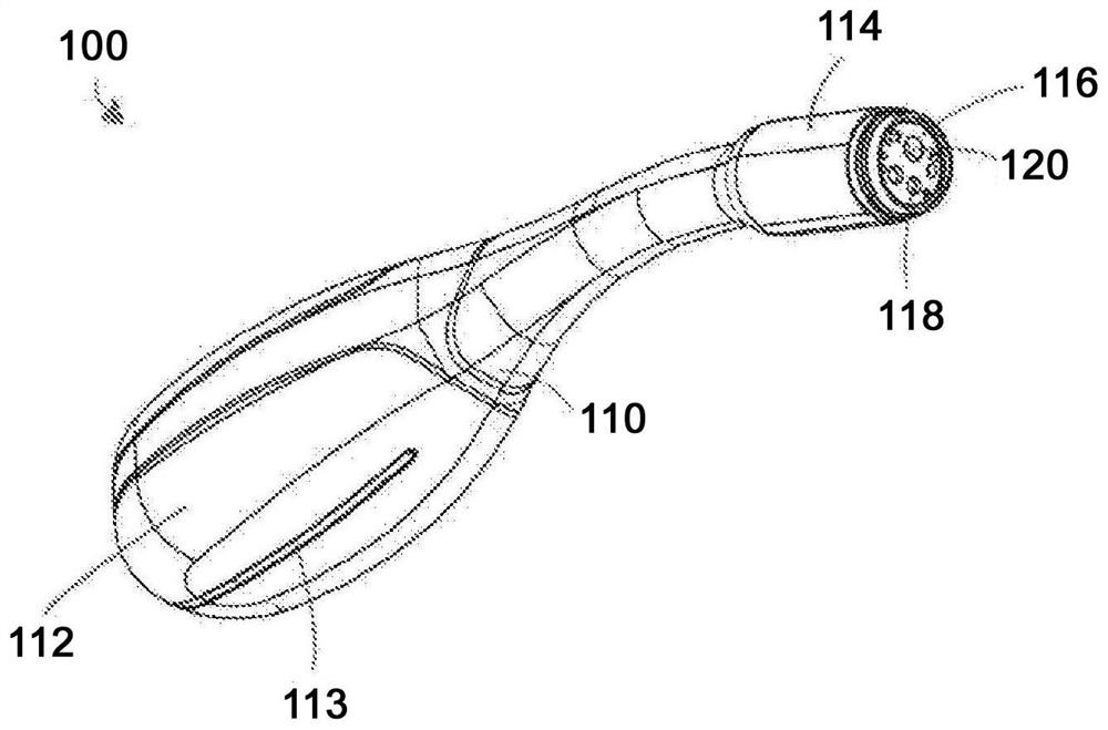
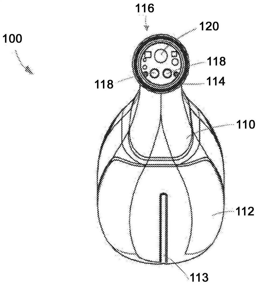
An imaging device includes a body having a first end portion configured to be held in a user's hand and a second end portion configured to direct light onto a surgical margin. The device includes at least one excitation light source configured to excite autofluorescence emissions of tissue cells and fluorescence emissions of induced porphyrins in tissue cells of the surgical margin. A white light source is configured to illuminate the surgical margin during white light imaging of the surgical margin. The device includes an imaging sensor, a first optical filter configured to permit passage of autofluorescence emissions of tissue cells and fluorescence emissions of the induced porphyrins in tissue cells to the imaging sensor, and a second optical filter configured to permit passage of white light emissions of tissues in the surgical margin to the imaging sensor. Systems and methods relate to imaging devices.

Description

[0001] Citations to related applications [0002] This application claims priority to U.S. Provisional Application No. 62 / 793,764 (filed January 17, 2019), entitled "Apparatus, System, and Method for Visualizing and Apparatus, System, and Method for Visualization and Removal of Tumors," U.S. Provisional Application No. 62 / 857,155 (filed June 4, 2019), the entire contents of which are incorporated herein by reference. technical field [0003] The present disclosure relates to devices, systems and methods for visualization and removal of tumors. The disclosed devices, systems, and methods can also be used to stage tumors and assess surgical margins, such as tissue margins on ex vivo tumors and tissue beds / surgical margins from which tumor and / or tissue have been removed edge of the bed. The disclosed devices, systems, and methods can also be used to identify one or more of residual cancer cells, precancerous cells, and satellite lesions, and provide guidance for removal and / or...

Claims


Application Information

Patent Timeline
Patent Type & Authority Applications(China)
IPC IPC(8): A61B5/00A61B46/10
CPCA61B46/10A61B5/0071A61B5/0091A61B90/39A61B90/37A61B10/0041A61B2090/3941A61B2090/3908A61B2090/373A61B2562/0242A61K49/0021A61K49/0034
Inventor 拉尔夫·达科斯塔凯瑟琳·奥托利诺-佩吕克里斯托弗·吉布森纳亚纳·塔兰基·阿南塔西蒙·特雷德韦尔托德·丹尼斯托德·米尼加雷特·弗迈卡尔·安妮斯
Owner SBI 艾拉制药加拿大有限公司
Why Eureka
Try Eureka