Tissue image recognition method and device, readable medium and electronic equipment

A technology for organizing images and recognition methods, applied in the field of image processing, can solve problems such as reducing the accuracy of image recognition and complicated work, and achieve the effect of improving practicability and accuracy

Active Publication Date: 2021-11-16
BEIJING BYTEDANCE NETWORK TECH CO LTD
View PDF9 Cites 2 Cited by
  • Summary
  • Abstract
  • Description
  • Claims
  • Application Information

AI Technical Summary

Problems solved by technology

However, labeling a large number of images requires a lot of manpower and material resources, and the work is compli

Method used


Image

  • Tissue image recognition method and device, readable medium and electronic equipment
  • Tissue image recognition method and device, readable medium and electronic equipment
  • Tissue image recognition method and device, readable medium and electronic equipment

Examples


Example Embodiment

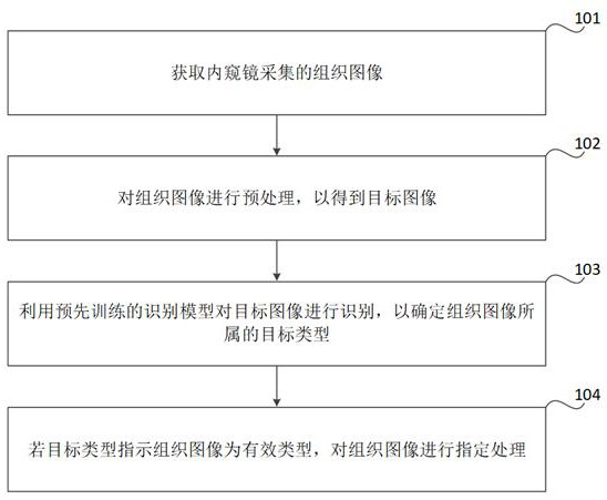
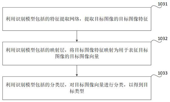
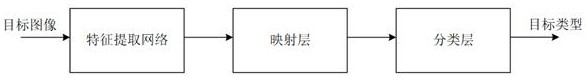
[0031] Embodiments of the present disclosure will be described in more detail below with reference to the accompanying drawings. Although certain embodiments of the present disclosure are shown, it is to be understood that the present disclosure can be implemented in various forms, and should not be construed as being limited to the embodiments set forth herein, and the opposite is provided for More thorough and completely understood. It should be understood that the accompanying drawings and examples of the present disclosure are for exemplary effects, not for limiting the scope of the present disclosure.

[0032] It should be understood that the various steps described in the method embodiments of the present disclosure can be performed in different order, and / or in parallel. Furthermore, the method embodiment may include additional steps and / or omitted the steps shown. The scope of this disclosure is not limited thereto.

[0033] The term "comprising" as used herein and its...


PUM


Abstract

The invention relates to a tissue image recognition method and device, a readable medium and electronic equipment, and relates to the technical field of image processing. The method comprises the steps: obtaining a tissue image collected by an endoscope, carrying out the preprocessing of the tissue image to obtain a target image, carrying out the recognition of the target image through a pre-trained recognition model, so as to determine the target type to which the tissue image belongs, wherein the recognition model is obtained through joint training with a preset comparison identification model according to a preset sample image set, the sample image set comprises a first number of labeled sample images with known types and a second number of unlabeled sample images with unknown types, and the first number is smaller than the second number; and comparing the structure of the recognition model to be the same as the structure of the recognition model, comparing neuron parameters of the recognition model, determining according to the neuron parameters of the recognition model, and if the target type indicates that the tissue image is an effective type, performing specified processing on the tissue image. The practicability and accuracy of the recognition model can be improved.

Description

technical field [0001] The present disclosure relates to the technical field of image processing, and in particular, to a tissue image recognition method, device, readable medium and electronic equipment. Background technique [0002] The endoscope is equipped with optical lens, image sensor, light source and other components, which can enter the inside of the human body for inspection, so that doctors can intuitively observe the internal conditions of the human body, and have been widely used in the medical field. In the process of using an endoscope for inspection, many invalid images may be collected due to unstable lens entry techniques or improper position of the endoscope, such as obstructions, overexposure, and sharpness. Too low etc. These invalid images will interfere with the inspection results of the endoscope. Therefore, it is necessary to identify the images collected by the endoscope to ensure the effectiveness and accuracy of the endoscope inspection. Usuall...

Claims


Application Information

Patent Timeline
IPC IPC(8): G06T7/00G06N3/04G06N3/08
CPCG06T7/0012G06N3/084G06T2207/10068G06T2207/20081G06T2207/20084G06N3/044G06N3/045
Inventor 边成杨志雄赵家英石小周李剑
Owner BEIJING BYTEDANCE NETWORK TECH CO LTD
Why Eureka
Try Eureka