Cone beam CT image reconstruction method
A CT image and cone beam technology, applied in the field of medical imaging, can solve the problems of image artifacts, image cupping artifacts, affecting image quality, etc., and achieve the effect of eliminating projection brightness deviation and reducing the impact.
- Summary
- Abstract
- Description
- Claims
- Application Information
AI Technical Summary
Problems solved by technology
Method used
Image
Examples
Embodiment 1
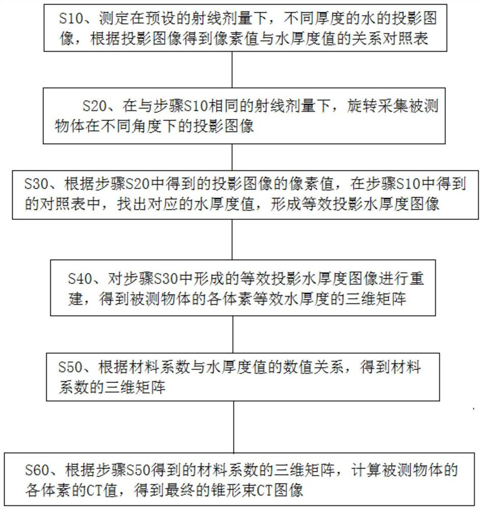
[0028] This embodiment provides a cone beam CT image reconstruction method for reducing the influence of scattered rays and ray hardening on cone beam CT image reconstruction results.
[0029] In the field of medical imaging technology, the CT value (unit: Hounsfield Unit, HU) of a spatial pixel (x, y, z) in a cone beam CT image can be calculated by the following formula:
[0030]
[0031] where μ Water is the X-ray linear attenuation coefficient of water at 73keV, in cm 2 / g; μ x (x, y, z) is the X-ray linear attenuation coefficient at a certain spatial pixel (x, y, z).
[0032] Thus, by obtaining μ x (x, y, z) and μ Water can calculate the CT value of a certain spatial pixel (x, y, z), and the cone beam CT image reconstruction can be regarded as solving μ x (x, y, z) process.
[0033] When performing cone beam CT image acquisition, the projection image can be regarded as the X-ray intensity distribution after passing through the measured object. Specifically, the X...
PUM
 Abstract
Description
Claims
Application Information
 - R&D
- Intellectual Property
- Life Sciences
- Materials
- Tech Scout
- Unparalleled Data Quality
- Higher Quality Content
- 60% Fewer Hallucinations
Browse by: Latest US Patents, China's latest patents, Technical Efficacy Thesaurus, Application Domain, Technology Topic, Popular Technical Reports.
© 2025 PatSnap. All rights reserved.Legal|Privacy policy|Modern Slavery Act Transparency Statement|Sitemap|About US| Contact US: help@patsnap.com


