

Application of miR-663a or target gene CKDN2A thereof in thin endometrium
A technology of mir-663a and endometrium, applied in the field of biomedicine, can solve problems such as decrease, increase, and unclear molecular mechanism
- Summary
- Abstract
- Description
- Claims
- Application Information
AI Technical Summary
Benefits of technology
Problems solved by technology
Method used
Image

Examples
Embodiment 1
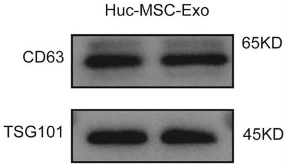
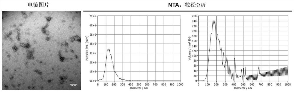
[0049] Example 1 Extraction and identification of HucMSC-EX
[0050] Extraction of exosomes from human umbilical cord mesenchymal stem cells (HucMSC-EX): clean bench operation, take the cell culture medium, divide into sterilized centrifuge bottles or centrifuge tubes, centrifuge at 4°C, 1000 x g for 10 min, remove Cell culture supernatant, debris and dead cells in culture medium;
[0051] Then, operate on an ultra-clean bench, filter the supernatant through a 0.2mm filter, and divide into sterilized centrifuge bottles; centrifuge the supernatant culture medium in an ultra-high-speed centrifuge at 4°C, 100,000 x g for 3 hours; discard the supernatant and add 10ml PBS, ultracentrifuge, 4°C, 100,000x g for 90min; discard the PBS, resuspend the exosome-containing particles at the bottom of the tube with 50μl PBS, transfer to a sterile imported 1.5ml EP tube, seal with parafilm, and transfer to Store at -80°C.
[0052] Identification of HucMSC-EX: HucMSC-EX were identified by We...
Embodiment 2
[0054] Example 2 Construction of EEC hypoxia model
[0055] (1) Human endometrial glandular epithelial cells (EEC) were cultured under normoxic conditions, and the cells were grown to a density / plate rate of about 60-70%.
[0056] (2) During hypoxia treatment, cells were exposed to humidified hypoxic air (1% O 2 ,94%N 2 ,5%CO 2 ), placed in an incubator (Thermo), and cultured at 37°C for 1 h, 4 h, 8 h, 16 h and 24 h; EECs cultured under normoxia were used as the control group.
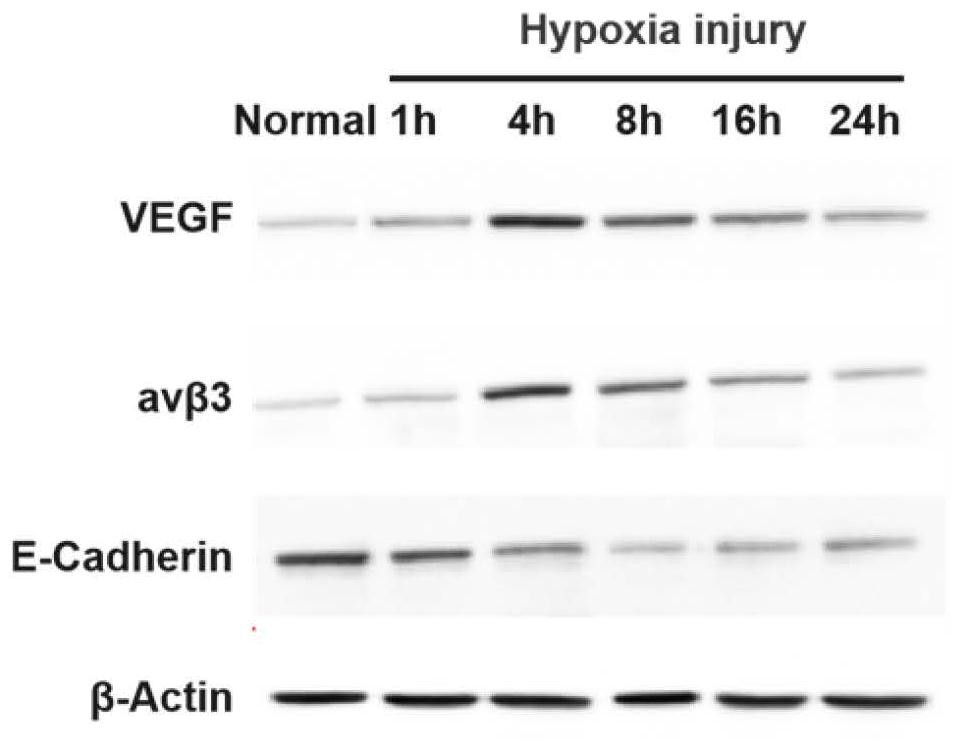
[0057] (3) Western blot was used to detect the expressions of VEGF (Abcam ab214424), avβ3 (Abcam ab179473) and E-Cadherin (Abcam ab40772) to identify hypoxic injury.
[0058] The result is as image 3 The results of Western Blot showed that after 4 hours of hypoxia, the hypoxia-related indicators VEGF and avβ3 were significantly up-regulated, and the expression of epithelial cell marker molecule E-Cadherin was also significantly down-regulated. Co-culture experiments were carried out.
Embodiment 3
[0059] Example 3 Hypoxic EEC to HucMSC exosome uptake experiment
[0060] HucMSC-Ex were co-cultured with EECs injured by hypoxia for 4 h and EECs treated with non-hypoxia, respectively, and then the two groups of cells were subjected to RKH67 fluorescence staining and observed under a 400-fold lens.
[0061] The specific steps are as follows. Exosome exosomes are labeled with PKH67 (company: Sigma-Aldrich, product number: MINI67-1KT). PKH67-labeled exosomes were co-cultured and incubated with HucMSCs for 24 hours; cells were fixed with paraformaldehyde (4%) for 15 minutes at room temperature; cells were punched with TBST in 0.5% Triton X-100 for 15 minutes at room temperature; labeled with DAPI staining Nuclei (4′,6-diamidino-2-phenylindole (DAPI) solution (Selleck S9980)); cellular uptake of labeled exosomes was detected using a laser scanning confocal microscope (OLYMPUS FLUOVIEW FV3000).
[0062] The result is as Figure 4 As shown, the results showed that the ability of...
PUM
| Property | Measurement | Unit |
|---|---|---|
| The peak | aaaaa | aaaaa |
Abstract
Description
Claims
Application Information