Weight used for partially scanning for quantum segmental multi-sectional CT imaging

A technique of CT scanning, pitch, used in the direction of instruments used for radiological diagnosis, computerized tomography scanners, generation of 2D images

Inactive Publication Date: 2005-09-07
GE MEDICAL SYST GLOBAL TECH CO LLC
View PDF0 Cites 0 Cited by
  • Summary
  • Abstract
  • Description
  • Claims
  • Application Information

AI Technical Summary

Problems solved by technology

Even if there is enough instantaneous resolution to allow seeking a minimum range of source angles in 180+ sector angles (π+2Γ), it is not always possible to achieve this minimum due to scanner speed and patient heart rate constraints

Method used


Image

  • Weight used for partially scanning for quantum segmental multi-sectional CT imaging
  • Weight used for partially scanning for quantum segmental multi-sectional CT imaging
  • Weight used for partially scanning for quantum segmental multi-sectional CT imaging

Examples


Embodiment Construction

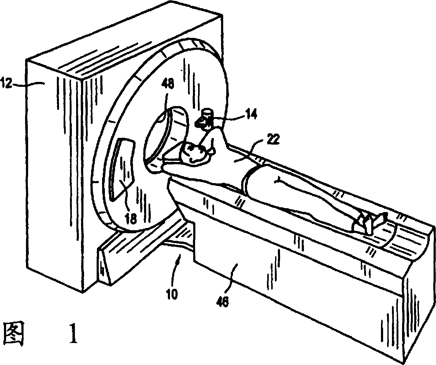
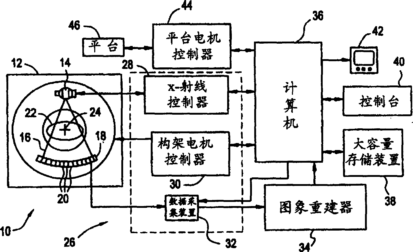
[0024] Referring to Figure 1 and figure 2 A computed tomography (CT) imaging apparatus 10 includes a gantry 12 representing a "third generation" CT scanner. The gantry 12 contains an x-ray source 14 which emits a beam of x-rays 16 towards a detector array 18 on the gantry 12 on the opposite side thereof. The detector array 18 consists of detector units 20 which detect X-rays emitted after passing through an object 22, such as a medical patient. The detector array 18 can be assembled in a single-slice or multi-slice configuration. Each detector unit 20 produces an electrical signal indicative of the beam-attenuated intensity of the active X-ray beam after passing through the patient 22 . During a scan in which X-ray projection data is collected, the gantry 12 and the components fixed thereto rotate about a center of rotation 24 .

[0025] The rotation of the gantry 12 and the operation of the X-ray source are managed by a control mechanism 26 of the CT apparatus 10 . The co...


PUM


Abstract

In one embodiment, the present invention is a method for reconstructing an image from a set of projection data acquired in a computed tomgraphic (CT) scan of an object (22). The method includes the steps of: selecting a pitch in a range between a zero pitch and a high-speed (HS) pitch; helically scanning the object with a CT imaging system (10) having a multislice detector (18) and a moving radiation source (14), at the selected pitch, to acquire projection data; helically interpolating the acquired projection data; to generate or synthesize projections in a plane of reconstruction for a source angle beta spanning a partial scan angle; applying a partial scan weight WPS written as: WPS( beta , gamma ) = beta -< pi >/2+ GAMMA /2( GAMMA - gamma )    beta inf = < pi >/2- GAMMA ≤ beta ≤ beta <-> = < pi >/2+ GAMMA -2 gamma WPS( beta , gamma ) = 1.0    beta <->≤ beta ≤ beta <+> = 3< pi >/2- GAMMA -2 gamma WPS( beta , gamma ) = 3< pi > /2+ GAMMA - beta /2( GAMMA + gamma )    beta <+>≤ beta ≤ beta sup = 3< pi >/2+ GAMMA to the generated or synthesized projections in a plane of reconstruction, where gamma is a fan angle, beta a source angle, and GAMMA a maximum fan angle; and filtering and backprojecting the weighted projections.

Description

technical field [0001] The present invention relates to a computerized tomography (CT) imaging method and its device, in particular to a method and its device for reconstructing an object image based on partial scanning. Background technique [0002] In at least one known computed tomography (CT) imaging apparatus, an X-ray source projects a fan-shaped beam that is collimated to lie in the X-Y plane of a Cartesian coordinate system, commonly referred to as the "imaging plane". flat". X-rays pass through the object to be imaged, such as a patient. The beam attenuated by the object impinges on the radiation detector array. The radiation intensity of the attenuated beam received by the detector array depends on how attenuated the X-rays are by the object. Each detector element in the detector array produces a separate electrical signal at the detector location which measures the degree of beam attenuation. Individual attenuation measurements are collected from all detectors...

Claims


Application Information

Patent Timeline
Patent Type & Authority Patents(China)
IPC IPC(8): A61B6/03G06T11/00
CPCY10S378/901G06T11/005G06T2211/421A61B6/027
Inventor G·M·贝森
Owner GE MEDICAL SYST GLOBAL TECH CO LLC
Why Eureka
Try Eureka