Method and apparatus for the preparation of a renal image

An image and kidney technology, applied in the medical field, can solve problems such as difficult image reprocessing and difficult to obtain correct images

Inactive Publication Date: 2006-10-18
GENERAL ELECTRIC CO
View PDF1 Cites 8 Cited by
  • Summary
  • Abstract
  • Description
  • Claims
  • Application Information

AI Technical Summary

Problems solved by technology

If a physician is required to perform a fairly complex operation, it will be difficult to ask him or her to reprocess the image with the same parameters as earlier
Without doing this, the two images are not really comparable, so it's hard to get a correct image of the effect of the processing described and what has been applied at the same time

Method used


Image

  • Method and apparatus for the preparation of a renal image
  • Method and apparatus for the preparation of a renal image

Examples


Embodiment Construction

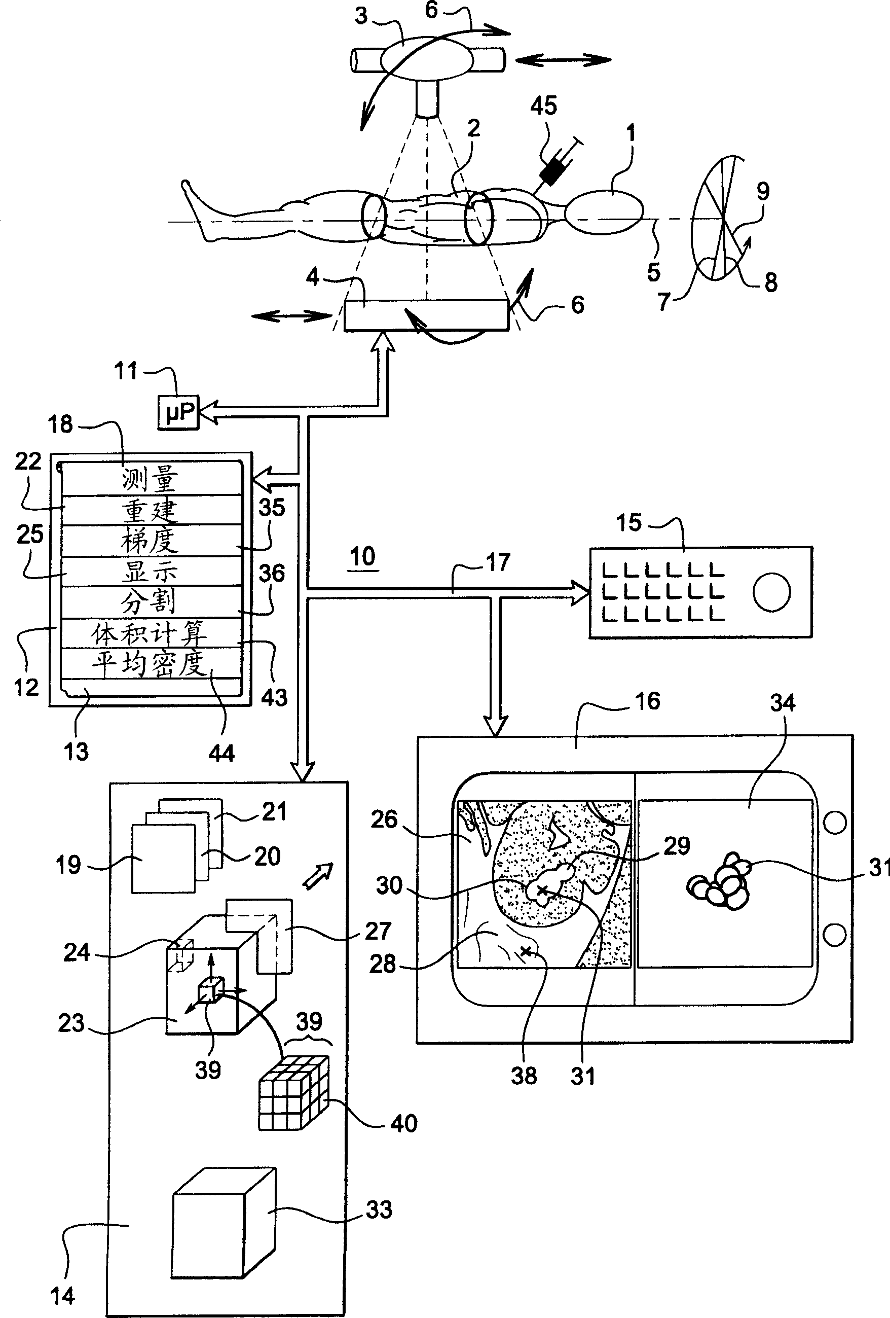
[0017] figure 1 An example of a conventional apparatus for carrying out method embodiments of the present invention is shown. In this method and apparatus, a subject's body 1 is subjected to a radiological examination. The body of the subject may be a human body, the kidneys of which are located in region 2 of the body. Region 2 of the body is the region to be examined. For example, the examination device comprises a computed tomography scanner schematically having an x-ray tube 3 and a detector 4 which together make a stepwise or continuous rotational movement 6 about an axis of rotation 5 . During said rotation, area 2 is subjected to a radiation sequence of X-rays irradiated from various angles, such as 7 to 9 . In computed tomography, for a given measurement accuracy, there are constraints on the angular distance of the different incident values ​​to which the body is irradiated and the minimum value of the detection angle (usually 180°). The apparatus is driven by a c...


PUM


Abstract

In an image of lithiasis in a computed tomography examination, a segment of the image is chosen with a detection of the maximum of the gradient of radiological density measured in the voxels of the reconstructed image. This mode of action gives an evaluation of the envelope of the lithiasis that is objective and independent of the practitioner who performs it. Furthermore, to make the characterization of the lithiasis more realistic, especially to follow the effects of a therapeutic treatment, a characteristic result is given, equivalent to the volume of the lithiasis, preferably a weighted volume, and to the mean density of the lithiasis. The disclosed method also can facilitate the scheduling of surgical operations when it is not possible to administer oral treatment or carry out lithotripsy.

Description

[0001] Cross References to Related Applications [0002] This application claims priority under 35 USC 119(a)-(d) to French Patent Application No. 0550218 filed January 25, 2005, the entire contents of which are hereby incorporated by reference. technical field [0003] One embodiment of the invention is a method and apparatus for preparing an image of a kidney, in particular by using a computed tomography scanner. The field of the invention is the medical field. Embodiments of the present invention are intended to facilitate the detection of the presence of calculus or calcifications also referred to as calculi or stones located within the body of a subject, such as a patient's kidneys, urinary organs (bladder, urethra, etc.) or gallbladder. Embodiments of the present invention also enable appropriate treatment of calculus and, in the case of surgery or procedures, provide tools for planning of surgery by computed tomography. Embodiments of the present invention also enable...

Claims


Application Information

Patent Timeline
Patent Type & Authority Applications(China)
IPC IPC(8): A61B6/03A61B6/00G06T7/00G06T15/08
CPCG06T7/0012A61B6/12G06T7/11G06T7/187G06T7/62G06T2200/24G06T2207/10081G06T2207/30084
Inventor M·D·费朗C·E·科里J·克诺普利奥奇J·-M·特勒特奈尔
Owner GENERAL ELECTRIC CO
Why Eureka
Try Eureka