

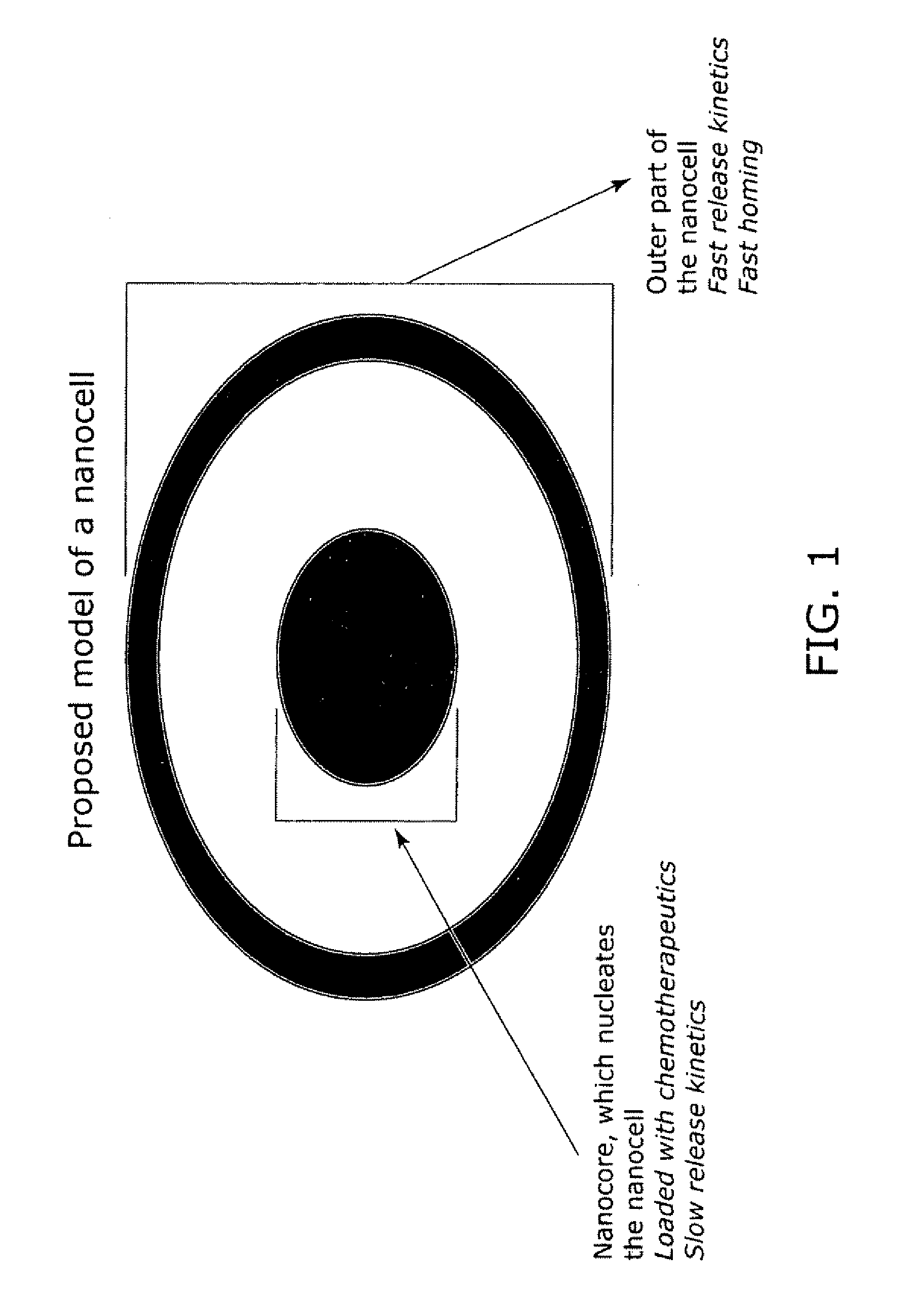
Nanocell drug delivery system
a technology of nanocell and drug delivery system, which is applied in the direction of aerosol delivery, powder delivery, granular delivery, etc., can solve the problems of patient suffering from the side effects of the anti-neoplastic agent without receiving any of its benefits, and collapse of the vasculature feeding the tumor,
- Summary
- Abstract
- Description
- Claims
- Application Information
AI Technical Summary
Benefits of technology
Problems solved by technology
Method used
Image

Examples
example 1
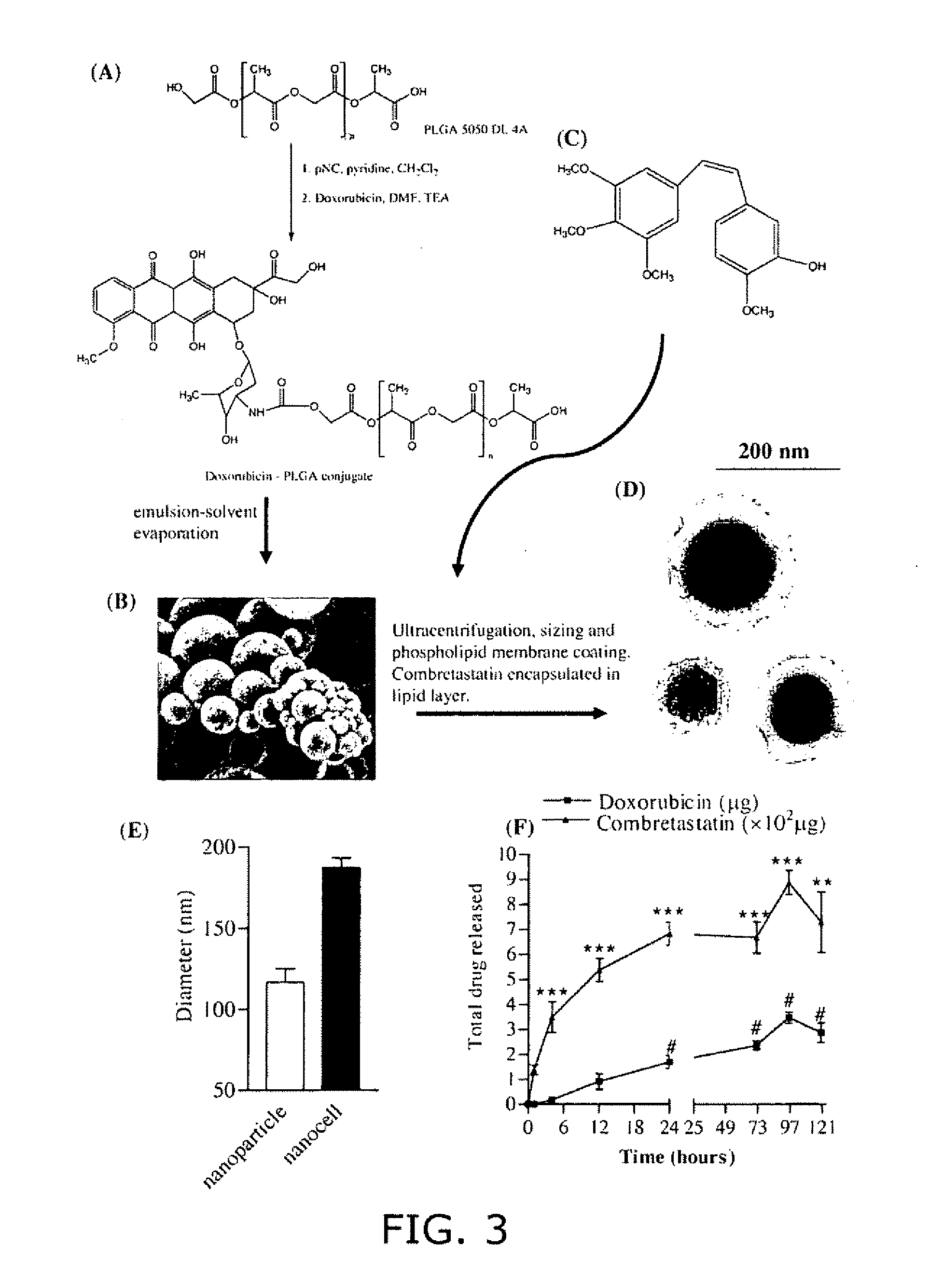
Synthesis and Analysis of Nanocells
[0180](A) Conjugation of Doxorubicin to PLGA (FIG. 3). Polylactic glycolic acid (PLGA) (Medisorb® 5050 DL 4A), having a lactide / glycolide molar ratio of 50 / 50, was obtained from Alkermes (Wilmington, Ohio). The average molecular weight of this polymer is reported to be 61 kDa, and it has free hydroxyl and carboxylic groups at its terminal ends. Doxorubicin hydrochloride, p-nitrophenyl chloroformate, and triethylamine were obtained from Sigma-Aldrich (St. Louis, Mo.). Briefly, 1.5 g of PLGA 5050 DL 4A was dissolved in 15 ml of methylene chloride and activated by the addition of 14 mg of p-nitrophenyl chloroformate and 9.4 mg (˜9.6 μl) of pyridine to the solution, kept in an ice bath at 0° C. (stoichiometric molar ratio of PLGA: p-nitrophenyl chloroformate: pyridine=1:2.8:4.7). The reaction was carried out for 3 hours at room temperature under nitrogen atmosphere. The resulting solution was diluted with methylene chloride and washed with 0.1% HCl and...
example 2
Developing the Novel In Vitro Assay System
[0189]Protocol: For setting up the system, human umbilical vein endothelial cells, pooled from three donors, were purchased from Cambrex, and used between passages 3-6. The cells were grown in endothelial basal medium supplemented with 20% fetal bovine serum (FBS) and bulletkit-2 (Sengupta et al. Cancer Res. 63(23):8351-59, Dec. 1, 2003). For the tumor component, we used Bl6 / F10 melanoma cells as the model cell line, which were stably transfected to express green fluorescent protein. Plasmid expressing enhanced green fluorescent protein (pEGFP-C2, Clontech) was linearized and lipofected (Lipofectamine 2000, Invitrogen) into Bl6-F10 cells. The stably integrated clones of Bl6-F10 cells were selected by 800 μg / ml G418. The green fluorescence of the G418 resistant clones was further confirmed by Flow Cytometry and epifluorescence microscopy. The GFP-Bl6 / F10 cells were regularly cultured in DMEM supplemented with 5% FBS. Sterile glass coverslips ...
example 3
In Vitro Efficacy of Drug Loaded Nanocells (FIG. 9)
[0194]Sterile glass coverslips (Corning) were coated with matrigel (extracellular matrix extracted from murine Englebreth-Holms sarcoma, diluted 1:3 in phosphate buffer saline; Becton Dickinson) or collagen (type I from rat's tail, Becton Dickinson). Synchronized human umbilical vein endothelial cells were trypsinised and plated on the coverslips at a density of 2×10⁴ cells per well. The cells were allowed to adhere for 24 hours in endothelial basal media supplemented with 20% fetal bovine serum. At this time point, the media was replaced with EBM supplemented with 1% serum, and green fluorescent protein-expressing Bl6 / F10 cells were added to the system at a density of 5×10³ cells per well. The co-culture was allowed to incubate overnight, following which different treatments were added to the media. At 24 hours post-treatment, the cells were fixed in paraformaldehyde (4% on ice, for 20 min), and stained with propidium iodide. The c...
PUM
| Property | Measurement | Unit |
|---|---|---|
| diameter | aaaaa | aaaaa |
| diameter | aaaaa | aaaaa |
| diameter | aaaaa | aaaaa |
Abstract
Description
Claims
Application Information