
Augmented surgical reality environment

a virtual reality environment and augmented technology, applied in the field of surgical techniques, can solve the problems of limited ability of existing robotic surgical tools, limited ability to identify conditions or objects, and limited ability of existing minimally invasive and robotic surgical tools, including but not limited to endoscopes and displays

Active Publication Date: 2018-12-11
COVIDIEN LP
View PDF13 Cites 616 Cited by
  • Summary
  • Abstract
  • Description
  • Claims
  • Application Information

AI Technical Summary

Benefits of technology

Enables clinicians to identify and address adverse conditions such as tissue perfusion, artery location, and vessel sealing effectiveness, reducing the need for additional invasive procedures and improving surgical outcomes by providing enhanced real-time visualization.

Problems solved by technology

The small incisions have reduced patient discomfort and improved recovery times. The small incisions have also limited the visibility of internal organs, tissue, and other matter.
Existing endoscopes and displays, especially those used in surgical robotic systems, have had a limited ability to identify conditions or objects that are within the field of view of the camera but are not fully visible within the spectrum shown on the display.
For example, existing minimally invasive and robotic surgical tools, including but not limited to endoscopes and displays, have had a limited, if any, ability to identify tissue perfusion after resection, locate different sized arteries within tissue, measure the effectiveness of vessel sealing, identify diseased or dead tissue from a heat signature, verify appropriate functioning after a resection, distinguish between sensitive areas (such as the ureter) and surrounding matter (such as surrounding blood), and detecting super-small leaks that are not visible with current tests.
In some surgeries these checks were either not performed or more invasive and / or time consuming tests were performed to check for these and other conditions and objects.

Method used


Image

  • Augmented surgical reality environment
  • Augmented surgical reality environment
  • Augmented surgical reality environment

Examples


Embodiment Construction

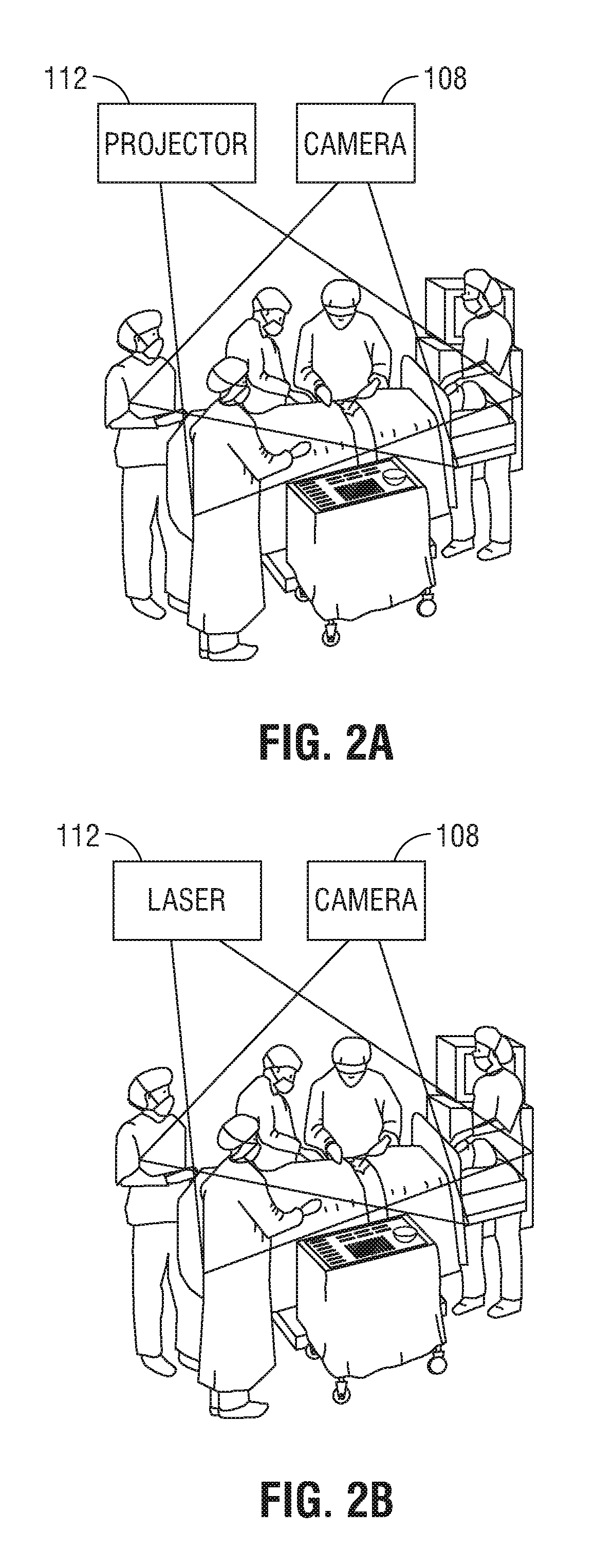
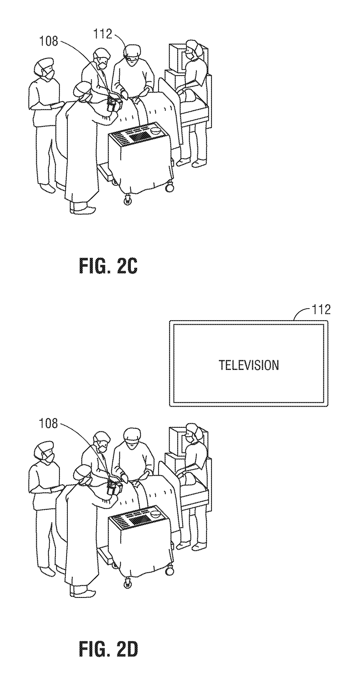
[0029]Image data captured from a surgical camera during a surgical procedure may be analyzed to identify additional imperceptible properties of objects within the camera field of view that may be invisible or visible but difficult to clearly see for people viewing the camera image displayed on a screen. Various image processing technologies may be applied to this image data to identify different conditions in the patient. For example, Eulerian image amplification techniques may be used to identify wavelength or “color” changes of light in different parts of a capture image. These changes may be further analyzed to identify re-perfusion, arterial flow, and / or vessel types.

[0030]Eulerian image amplification may also be used to make motion or movement between image frames more visible to a clinician. In some instances changes in a measured intensity of predetermined wavelengths of light between different image frames may be presented to a clinician to make the clinician more aware of t...


PUM


Abstract

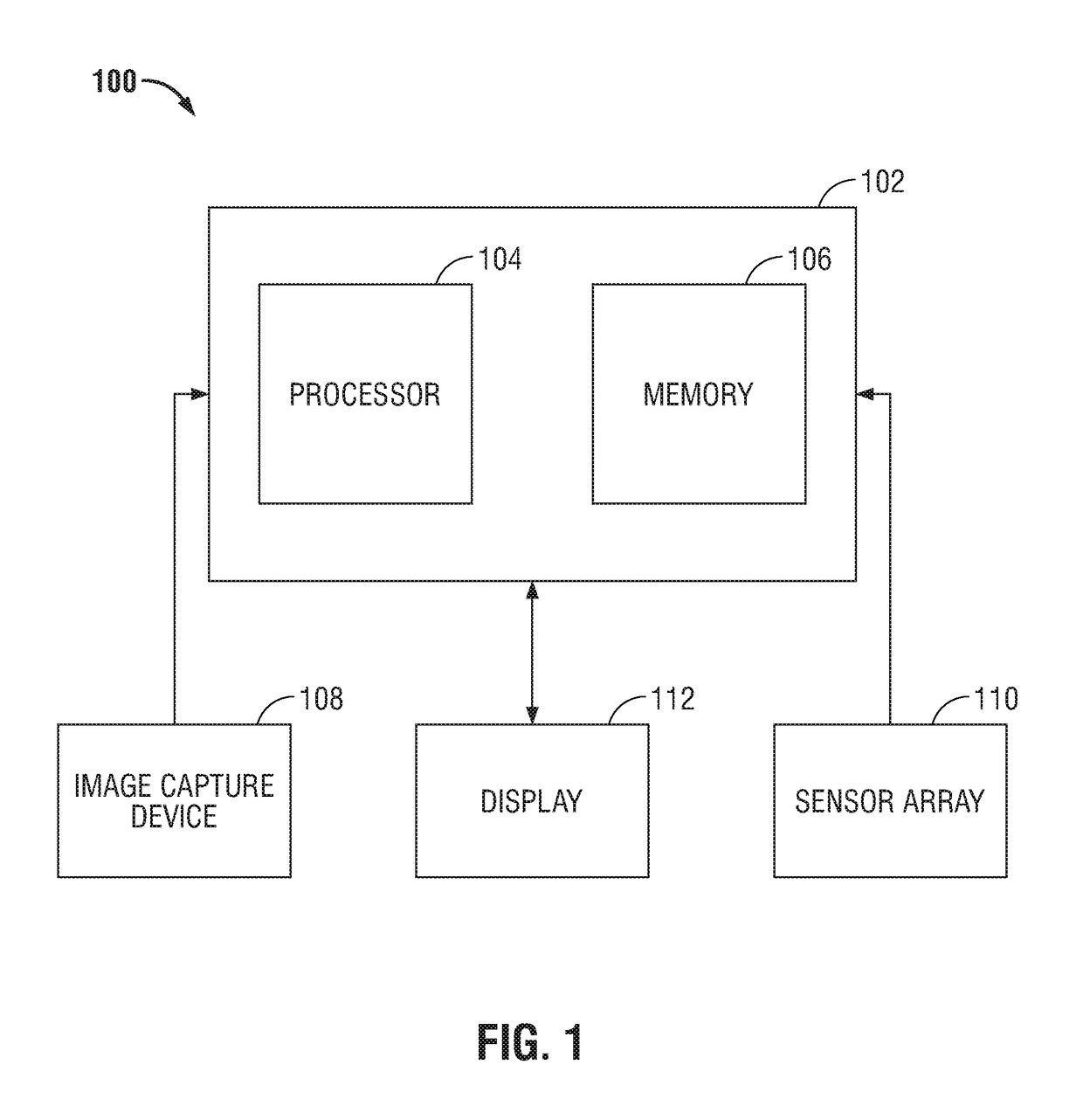
The present disclosure is directed to an augmented reality surgical system for viewing an augmented image of a region of interest during a surgical procedure. The system includes an image capture device that captures an image of the region of interest. A controller receives the image and applies at least one image processing filter to the image. The image processing filter includes a spatial decomposition filter that decomposes the image into spatial frequency bands. A temporal filter is applied to the spatial frequency bands to generate temporally filtered bands. An adder adds each band spatial frequency band to a corresponding temporally filtered band to generate augmented bands. A reconstruction filter generates an augmented image by collapsing the augmented bands. A display displays the augmented image to a user.

Description

CROSS REFERENCE TO RELATED APPLICATIONS[0001]This application is a U.S. National Stage Application filed under 35 U.S.C. § 371(a) of International Patent Application No. PCT / US2015 / 041083, filed Jul. 20, 2015, which claims the benefit to U.S. Provisional Patent Application No. 62 / 028,974, filed Jul. 25, 2014, the entire disclosure of each of which is incorporated by reference herein.BACKGROUND[0002]1. Technical Field[0003]The present disclosure relates to surgical techniques to improve surgical outcomes for a patient. More specifically, the present disclosure is directed to systems and methods for augmenting and enhancing a clinician's field of vision while performing a surgical technique.[0004]2. Background of the Related Art[0005]Minimally invasive surgeries have involved the use of multiple small incisions to perform a surgical procedure instead of one larger opening. The small incisions have reduced patient discomfort and improved recovery times. The small incisions have also li...

Claims


Application Information

Patent Timeline
Patent Type & Authority Patents(United States)
IPC IPC(8): G06T7/00H04N5/33A61B90/00G06T7/13G06T19/20G06T19/00G06T5/10G06T11/00H04N5/232
CPCG06T7/0012A61B90/37G06T5/10G06T7/13G06T11/008G06T19/006H04N5/232H04N5/33G06T19/20G06T2207/30004A61B2090/365A61B2576/00G06T2207/20024G06T5/50G06T2207/10048G06T2207/20221H04N23/63H04N23/20A61B90/361H04N23/60
Inventor CARNES, TONYMCKENNA, EDWARDPACK, STEPHEN
Owner COVIDIEN LP
Learn More