
Evaluation of an x-ray image of a breast produced during a mammography

a breast and x-ray technology, applied in the field of evaluating an x-ray image of a breast produced during a mammography, can solve the problems of increased risk of breast cancer for women whose breast has a high vbd value, insufficient use of vbd value for accurate description of masking, and risk of errors of judgment, so as to improve the accuracy of classification into breast density categories to be undertaken, reduce subjective influences and errors, and improve the effect of classification

Active Publication Date: 2019-01-01
SIEMENS HEALTHCARE GMBH
View PDF26 Cites 2 Cited by
  • Summary
  • Abstract
  • Description
  • Claims
  • Application Information

AI Technical Summary

Benefits of technology

The patent describes a method for automatically evaluating breast density by taking into account masking properties of fibroglandular tissue. This method allows for a more accurate and consistent evaluation of breast density compared to conventional methods, reducing subjective errors and simplifying the evaluation process. It provides a standardized method for categorizing breast density and meets the requirements of the 5th edition of BI-RADS. Overall, this method provides a more reliable and efficient tool for diagnostic breast imaging.

Problems solved by technology

Women whose breast has a high VBD value have an increased risk of getting breast cancer.
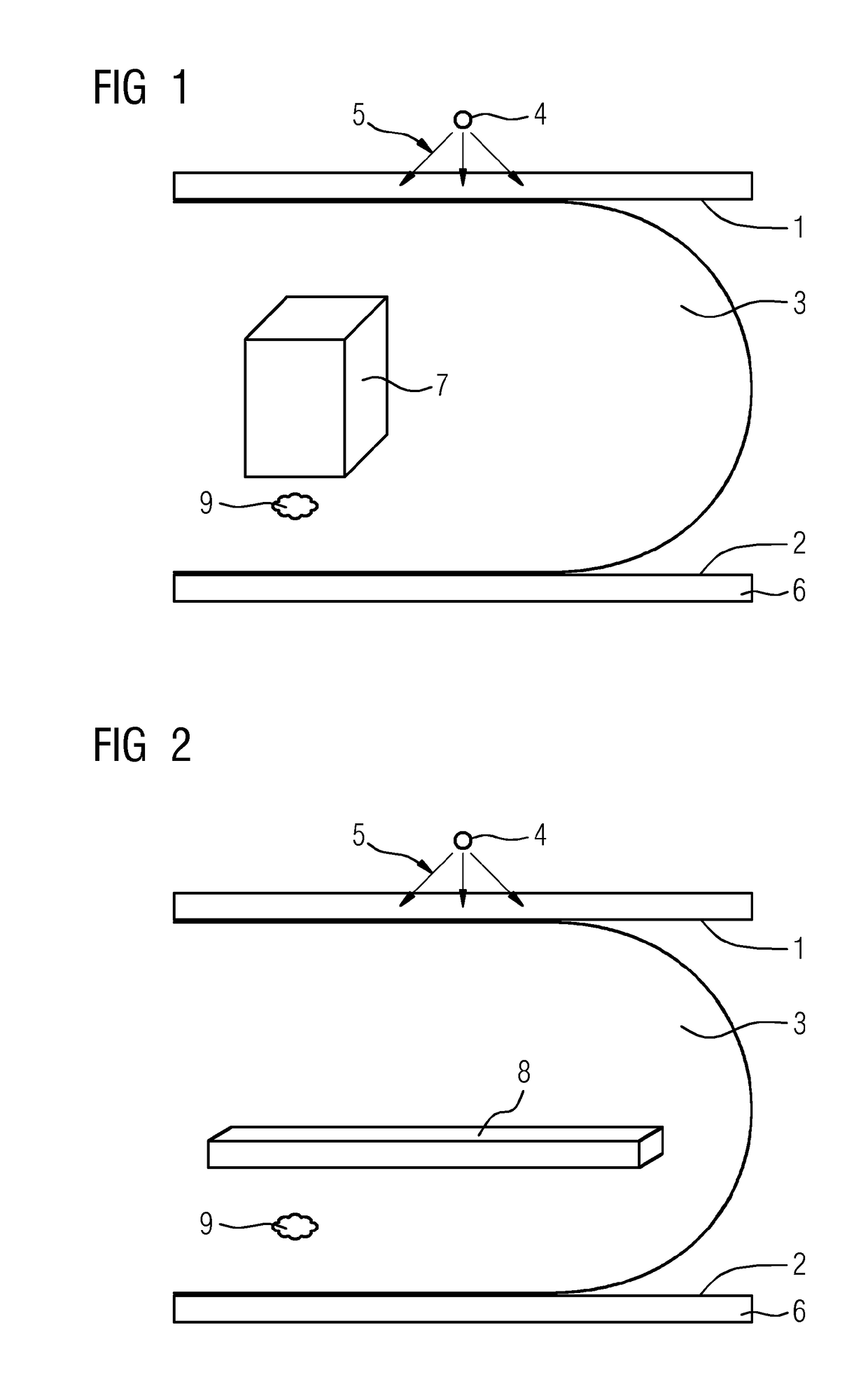
Therefore, the sole use of the VBD value is not sufficient for an accurate description of the masking effect by mammographically dense tissue 7, 8.
Here, there is a risk of errors of judgment.

Method used


Image

  • Evaluation of an x-ray image of a breast produced during a mammography
  • Evaluation of an x-ray image of a breast produced during a mammography
  • Evaluation of an x-ray image of a breast produced during a mammography

Examples


Embodiment Construction

[0030]The masking risk caused by mammographically dense tissue 7, 8 is determined automatically and used for categorizing, describing and / or representing the breast density in the method for evaluating an x-ray image 10 of a breast 3 produced during mammography. The x-ray image 10 to be evaluated may be a full-field mammography (FFDM) image or a projection image from a digital breast tomosynthesis (DBT) record.

[0031]If reference is made below to an “x-ray image”, this need not relate to the whole record. Instead, this may also only relate to a currently displayed or processed image region of the recorded x-ray image.

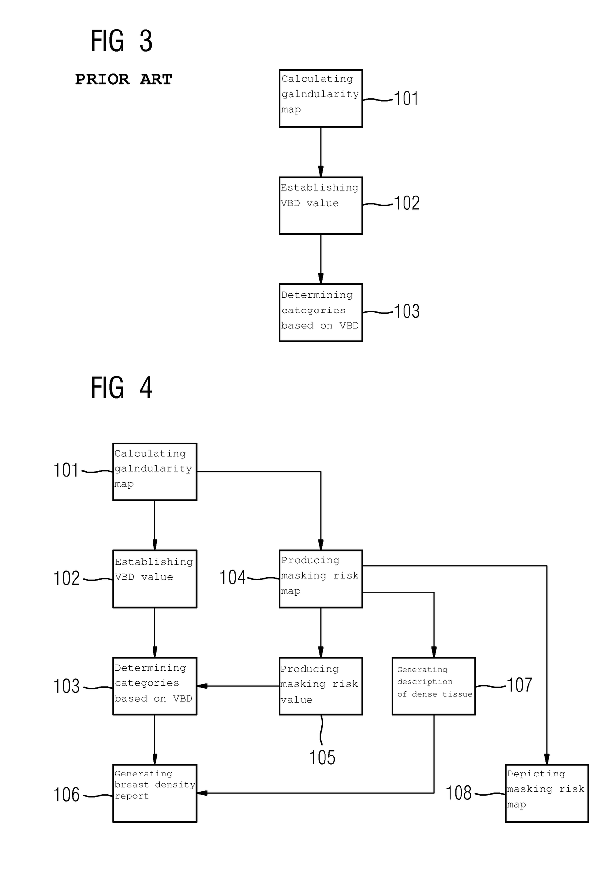
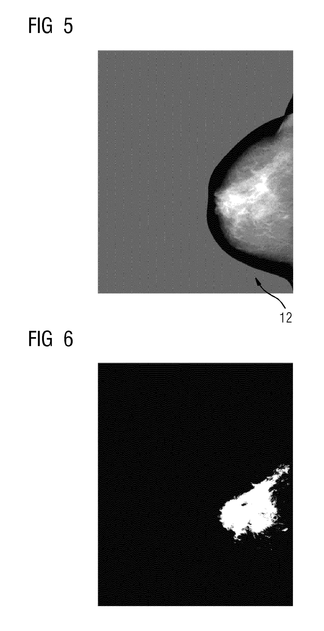
[0032]The basis of the method is the automatic production of a masking risk map 11 for the region of the breast 3 depicted in the x-ray image; see FIG. 4, act 104. This masking risk map 11 depicts the interconnected, dense tissue. Below, an exemplary way for producing such a masking risk map 11 is described.

[0033]The data from a glandularity map 12 (G(x,y)) as depicted i...


PUM


Abstract

The embodiments relate to a method, an apparatus, and a computer program for evaluating an x-ray image of a breast produced during a mammography. In order to simplify the evaluation of such an x-ray image in respect of the breast density, a method is proposed to automatically determine the masking risk caused by the mammographically dense tissue and to use this for categorizing, describing, and / or representing the breast density.

Description

[0001]The present patent document is a § 371 nationalization of PCT Application Serial Number PCT / EP2015 / 054429, filed Mar. 3, 2015, designating the United States, which is hereby incorporated by reference, and this patent document also claims the benefit of DE 10 2014 208 411.3, filed May 6, 2014, which is also hereby incorporated by reference.TECHNICAL FIELD[0002]The embodiments relate to a method, an apparatus, and a computer program for evaluating an x-ray image of a breast produced during mammography.BACKGROUND[0003]Automatically calculating the volumetric breast density (VBD) from an x-ray image produced during mammography is known. The volumetric breast density is defined as the ratio of the volume of the fibroglandular tissue to the overall volume of the breast. Below, the terms “fibroglandular tissue,”“glandular tissue,” and “mammographically dense tissue” or “dense tissue” are used synonymously. On the basis of this VBD value, the breast has until now been assigned a speci...

Claims


Application Information

Patent Timeline
Patent Type & Authority Patents(United States)
IPC IPC(8): G06T7/00A61B5/00
CPCG06T7/0012A61B5/4312G06T2207/30068G06T2207/20036G06T2207/10116
Inventor FIESELMANN, ANDREAS
Owner SIEMENS HEALTHCARE GMBH
Learn More