Method and probe for monitoring oxygen status in live mammalian cells

a live mammalian cell and oxygen status technology, applied in the field of methods and probes for monitoring oxygen status in live mammalian cells, can solve the problems of low loading efficiency, low loading efficiency, complex and invasive,

Active Publication Date: 2019-12-17
UNIV COLLEGE CORK NAT UNIV OF IRELAND CORK
View PDF3 Cites 0 Cited by
  • Summary
  • Abstract
  • Description
  • Claims
  • Application Information

AI Technical Summary

Benefits of technology

[0029]The method of the invention may be employed to monitor oxygen status in individual cells or cell populations (for example, cells grown in-vitro or ex-vivo cell or tissue samples). The method of the invention may also be employed to monitor cell health or cell function. For example, as oxygen levels or use can function as a diagnostic variable of cellular metabolism, the method of the invention can be employed to monitor metabolism in a cell, and the effects on metabolism caused by various compounds or conditions, for example drugs, chemicals, toxins, growth factors, hormones, low molecular weight effectors of gene transcription and translations, and environmental conditions. The fact that the probe employed in the present invention exhibits little or no intrinsic toxicity to cells allows for a more accurate assessment of cellular metabolism using the method of the invention. Indeed, it is another unexpected advantage of the present invention that high loading of probe (i.e. loading that enables stable signal intensity (see FIG. 3) and reliable lifetime-based sensing of intracellular oxygen) does not result in toxicity to the cell.
[0030]Various techniques are available for measuring the photoluminescent signal, including time-resolved fluorimetry in the microsecond domain and fluorescence microscopy imaging technique. Ideally, the photoluminescence lifetime of the photoluminescent signal, or parameter related to it, is measured, although the method of the invention may also employ signal phase or intensity measurements.
[0031]In one embodiment, the method of the invention also employs ratiometric intensity based oxygen sensing or imaging. In such embodiments, the photoluminescent probe comprises an additional oxygen-insensitive dye which is used as a reference or as part of a FRET pair. Examples of such dyes include oxygen-insensitive fluorescent dyes (i.e Oregon Green 488-dextran).
[0032]The invention also provides a method of determining the metabolic status of a cell or a cell population, which method comprises a step of monitoring oxygen status of the cell or cell population according to a method of the invention, and correlating the measured oxygen status, or changes in the oxygen status, of the cell or cell population to metabolic status of the cell or cell population. Generally, the method is used to monitor metabolic activity, often in response to a stimulus, for example a drug or effector or an environmental condition. Generally, the method involves monitoring oxygen concentrations, especially changes in oxygen concentration.
[0033]Thus, in one aspect, the invention provides a method of determining the effects of a test substance or condition on the metabolic activity of a cell or cell population, the method comprising a step of stimulating a cell or cell population with the test substance or condition, and determining the metabolic activity of the cell or cell population prior to and after stimulating the cell or cell population, according to the method of the invention.
[0034]The invention also relates to a method of determining oxygen concentration in a live mammalian cell sample by photoluminescence quenching technique, which method employs a photoluminsecent probe, the method comprising the step of:

Problems solved by technology

These probes were designed primarily for extracellular use, they are essentially cell-impermeable and can not be used for probing (intra)cellular O2.
However, these systems have low loading efficiency, uneven distribution of the probe inside the cell, uncontrolled compartmentation and aggregation, significant cyto- and phototoxicity, they are rather complex and invasive.
In many cases, such loading is low, cell-specific and stressful, time-consuming and not very reproducible.
Thus, the former probe has low brightness and short fluorescence lifetime resulting in modest sensitivity to O2, and their utility in biological applications has not been demonstrated.
The latter probes based on phosphorescent Pt- or Pd-coproporphyrins have modest photostability making them unusable in fluorescence oxygen imaging applications.
Although these probes are brighter than Ru(II)-probes (higher molar absorptivity and emission yield), not always they provide sufficient signals for reliable and accurate O2 sensing experiments.
Both probe types are limited to just a few dye structures, complex synthesis procedures and high costs.

Method used


Image

  • Method and probe for monitoring oxygen status in live mammalian cells
  • Method and probe for monitoring oxygen status in live mammalian cells
  • Method and probe for monitoring oxygen status in live mammalian cells

Examples


example 1

Fabrication of the Nanoparticle O2 Probe

[0065]1.5 g of RL-100 polymer and 22.5 mg of PtTFPP were dissolved in 750 g of acetone. The solution was placed in a 5 L beaker to which 4 L of deionized water were added over 20 s under rigorous stirring. Acetone was subsequently removed under reduced pressure and aqueous dispersion of the beads was further concentrated to approximately 75 mL. Traces of aggregates were removed by filtration through a paper filter. The resulting solution was filtered through a sterile filter to produce stock of O2 probe for intracellular use. It was aliquoted and stored in a dark place at +4° C. for several months for further use.

example 2

Assessment and Optimisation of Cell Loading

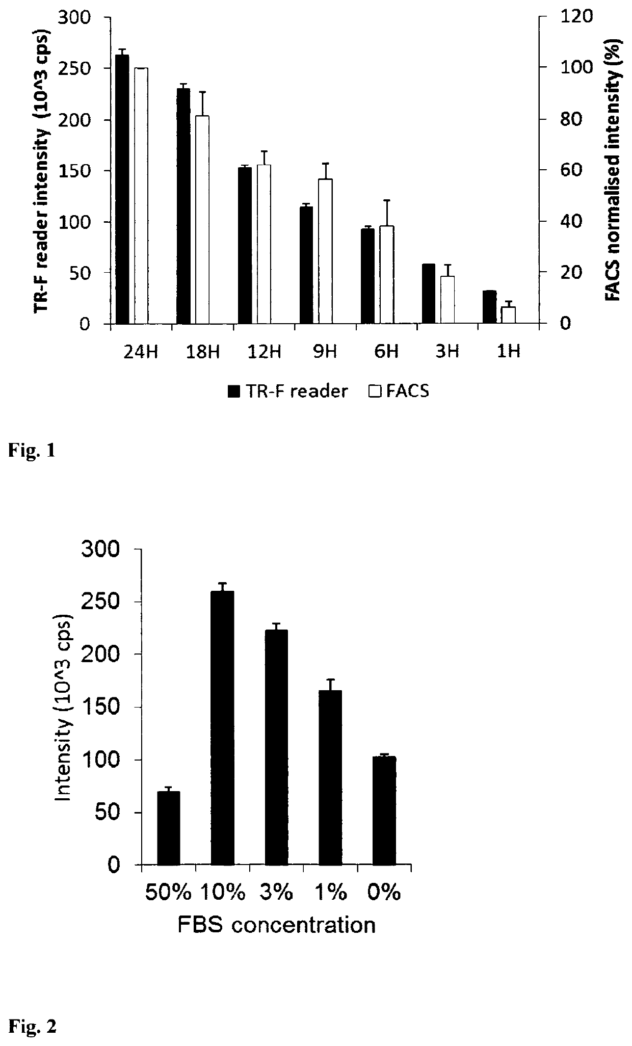
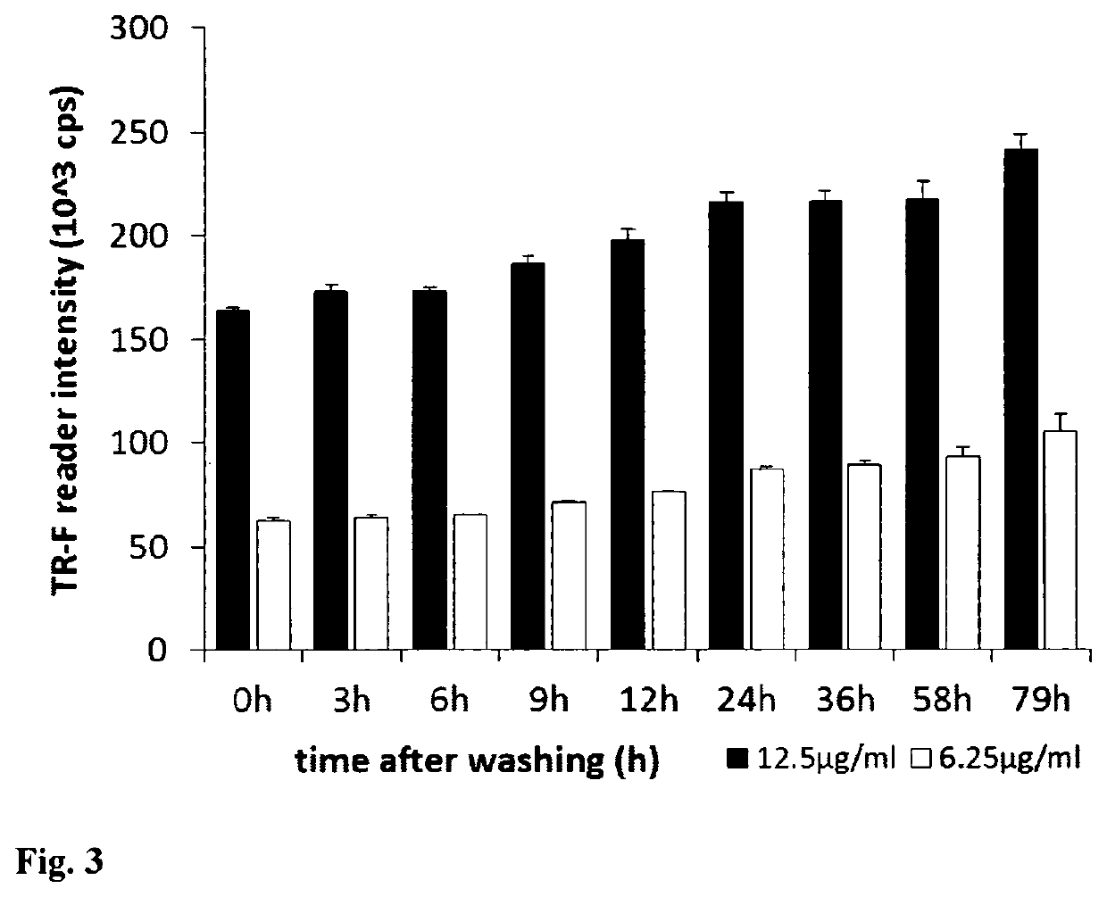
[0066]Mouse embryonic fibroblast (MEF) cells were cultured in 96-well plates using DMEM medium supplemented with 10% of foetal calf serum, CO2 incubator at 37° C., until they reach high confluence (periodically changing medium, if required). Stock solution of the PtPFPP-RL100 probe was diluted to the desired concentration with appropriate growth medium (determined by the cells and culturing conditions) and added to the wells with growing cells at the required final concentration (usually 1-10 μg / ml). Cells were incubated with probe, typically for 6-24 h, and then washed two times with fresh medium. Similar experiments were conducted with other cells, probe concentrations and incubation times. The efficiency of cell loading was assessed by measuring phosphorescence intensity signals in each well on a fluorescent reader Victor2 (Perkin Elmer) in time-resolved mode under the following settings: excitation −340 nm, emission −642 nm, delay time ...

example 3

Analysis of Probe Cytotoxicity and Localisation

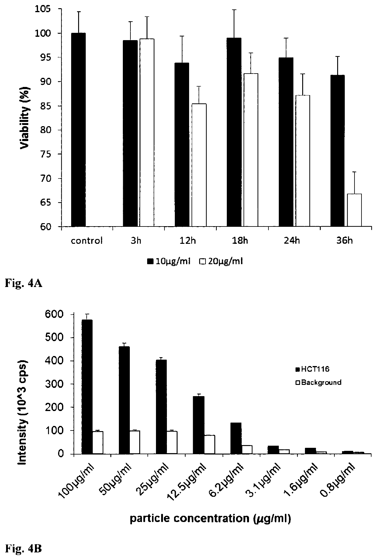
[0069]MEF, PC12, HepG2 and HCT116 cells were loaded with 10 μg / ml of PtPFPP-RL100 probe as described in Example 2. Probe cytotoxicity was assessed by measuring total ATP levels in loaded cells and comparing them with unloaded cells. FIG. 4A shows that the probe has low intrinsic toxicity, particularly when used at low concentrations (10 μg / ml) and short loading times (<18 h). FIG. 4B shows the concentration dependence of specific signal from HCT116 cells loaded with R1100-PtTFPP for 14h and background (non-specific binding to plate). Concentrations higher than 25 ug / ml had a toxic effect on cells.


PUM

PropertyMeasurementUnit
concentrationaaaaaaaaaa
sizeaaaaaaaaaa
concentrationaaaaaaaaaa

Abstract

The invention is based on the use of photoluminescent probes for intracellular sensing of oxygen, especially assaying intracellular oxygen concentration. The photoluminescent probe comprises a suspension of polymeric particles having an average diameter in the 20 nm to 100 nm range, formed from an amphiphilic cationic co-polymer which is oriented in the formed particle to provide a hydrophobic core and a hydrophilic shell. The probe includes a hydrophobic oxygen-sensitive photoluminescent dye such as Pt-tetrakis(pentafluorophenyl)porphine, PtPFPP embedded in the hydrophobic core of the particle, and the co-polymer includes quaternary ammonium groups which provide hydrophilic and cationic character to the particle shell. The photoluminescent probe, which in use is provided in the form of an aqueous suspension of probe, is incubated with live mammalian cells in a suitable growth medium for a period of time to allow the probe particles passively load into the cells. Oxygen can then be sensed by detecting a photoluminescent signal of the photoluminescent probe, which can be correlated with oxygen status, for example oxygen concentration or changes in oxygen concentration / levels, using existing techniques.

Description

CROSS REFERENCE TO RELATED APPLICATION[0001]This application is the national phase under 35 USC 371 of International Application No. PCT / EP2010 / 066006, filed Oct. 22, 2010. The content of the aforementioned application is incorporated herein in its entirety.INTRODUCTION[0002]The invention describes the use of photoluminescent probes for assaying oxygen levels in live mammalian cells. In particular, the invention describes photoluminescent oxygen-sensitive nanoparticles which have the ability to passively load various mammalian cells and report on local cellular oxygen levels by changing their phosphorescence characteristics.BACKGROUND TO THE INVENTION[0003]Quantification of O2 by photoluminescence quenching is an established technique which has a number of attractive features, including reversible, non-chemical and non-invasive nature of sensing of O2. This methodology is actively used in various industrial and biomedical applications where measurement of O2 is required (Papkovsky D...

Claims


Application Information

Patent Timeline
Patent Type & Authority Patents(United States)
IPC IPC(8): G01N21/64G01N33/50G01N33/84G01N33/58B82Y30/00
CPCB82Y30/00G01N33/587G01N33/84G01N21/6486G01N33/5008G01N33/5005G01N33/582
Inventor PAPKOVSKY, DMITRIFERCHER, ANDREASKLIMANT, INGOBORISOV, SERGEYZHDANOV, ALEXANDER
Owner UNIV COLLEGE CORK NAT UNIV OF IRELAND CORK
Why Eureka
Try Eureka