Processing at least one X-ray image

a technology of x-ray and image, applied in image data processing, instruments, computing, etc., can solve the problems of increasing noise level in images, accidentally removing structures within images, and lowering the dosage, so as to reduce noise and save structure

US10832381B2Active Publication Date: 2020-11-10SIEMENS HEALTHCARE GMBH
6 Cites 0 Cited by

Patent Information

Authority / Receiving Office
US · United States
Patent Type
Patents(United States)
Current Assignee / Owner
Publication Date
2020-11-10

Smart Images

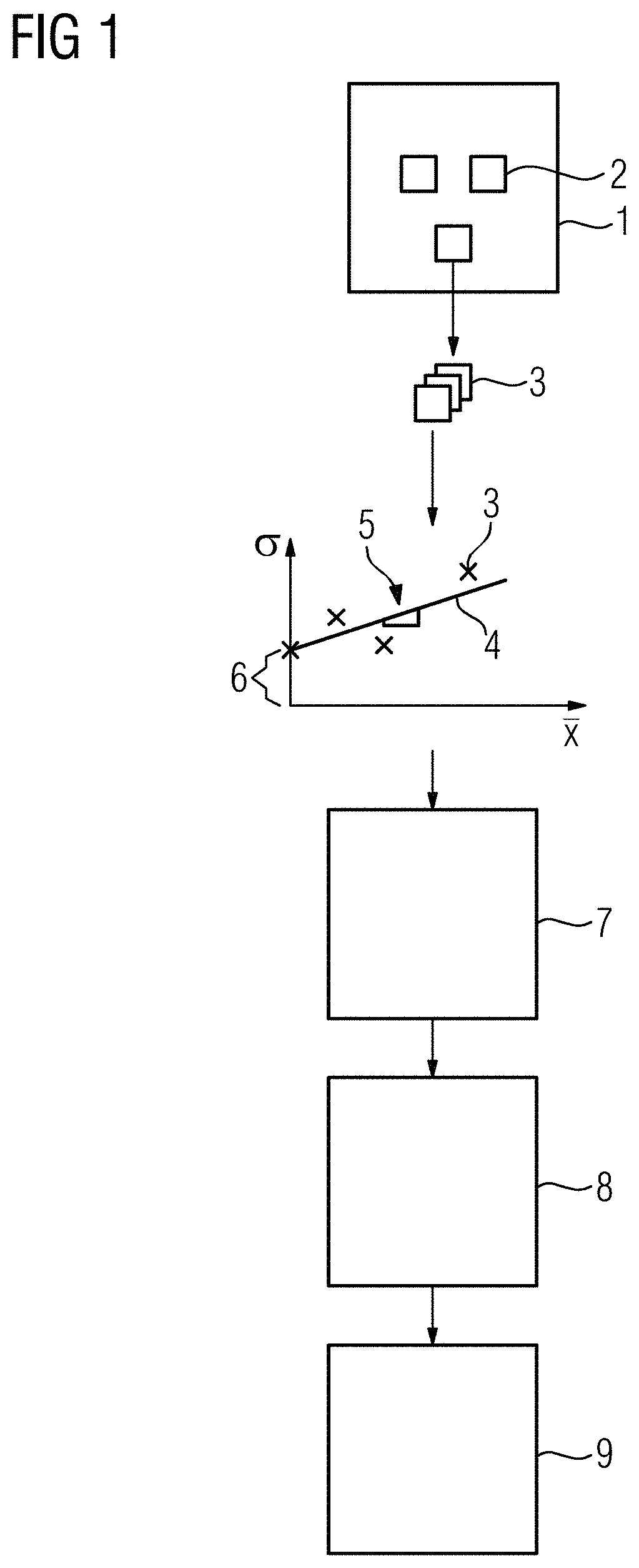
  • Figure 1
    Figure 1
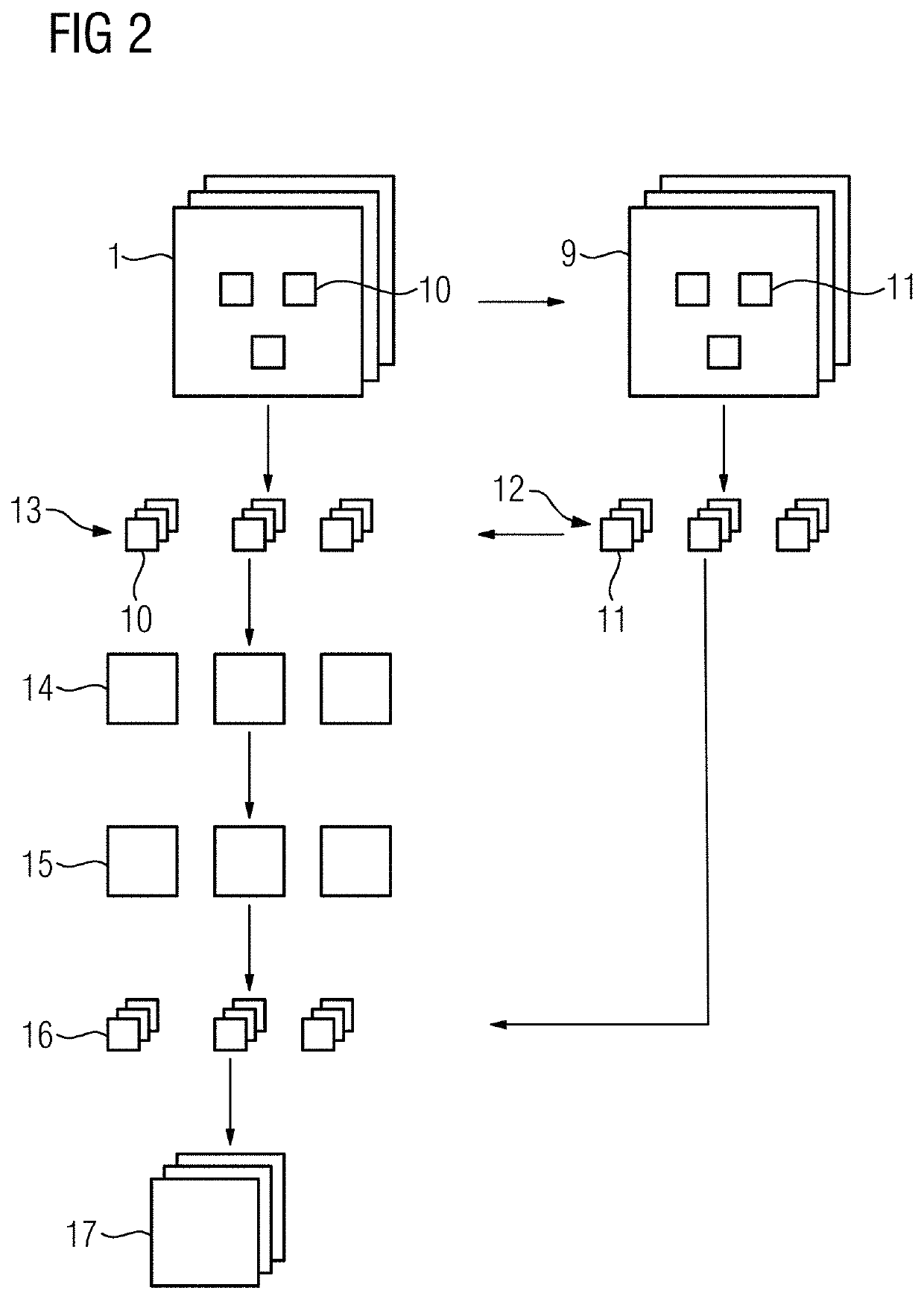
  • Figure 2
    Figure 2
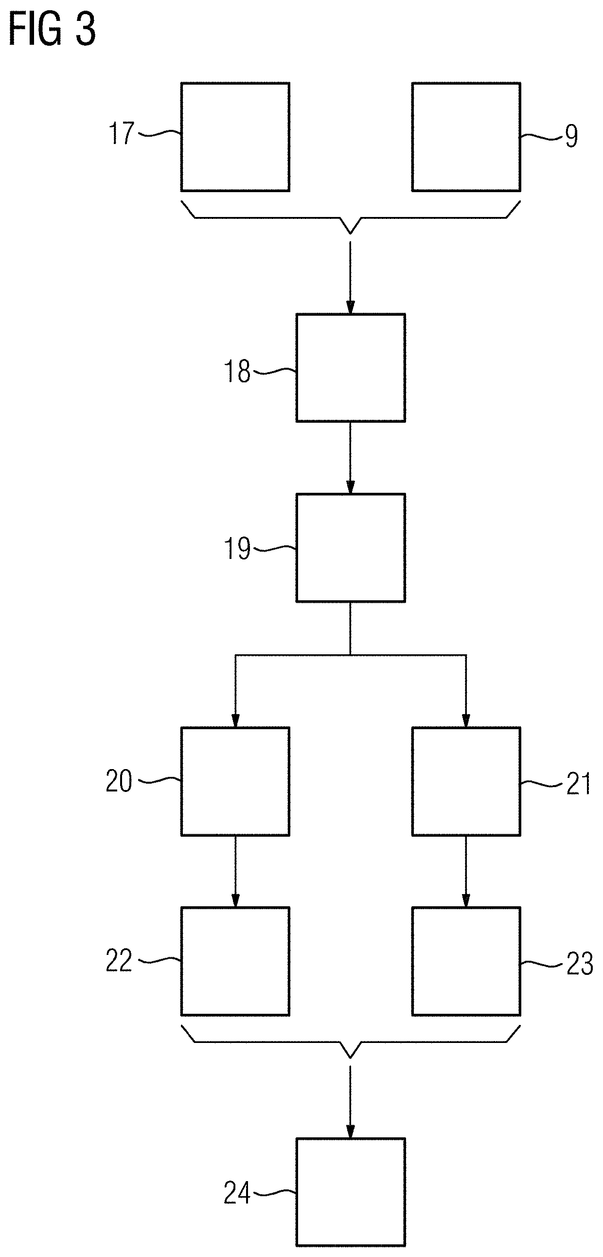
  • Figure 3
    Figure 3
Patent Text Reader

Abstract

A method for processing at least one X-ray image is provided. A variance of noise is signal dependent. The method includes applying a variance-stabilizing transformation to image data of the at least X-ray image to generate variance-stabilized data. At least one transform parameter of the variance-stabilizing transformation is dependent on a property of the at least one X-ray image that depends on an X-ray imaging device and / or a measurement parameter used to record the at least one X-ray image. A noise reduction algorithm is applied to the variance-stabilized data to generate noise-reduced data, and an inverse transformation of the variance stabilizing transformation is applied to the noise-reduced data to generate a denoised X-ray image.
Need to check novelty before this filing date? Find Prior Art

Description

[0001] This application claims the benefit of EP17183525.9, filed on Jul. 27, 2017, which is hereby incorporated by reference in its entirety.BACKGROUND

[0002] The present embodiments are related to processing at least one X-ray image.

[0003] In X-ray imaging (e.g., in medical imaging), it is desirable to use low X-ray intensities to lower the X-ray dose radiated onto the subject. Reducing the intensity of the X-ray radiation and / or shortening the acquisition time will lower the dosage, but at the same time, the noise level in the images will increase. The noise in X-ray imaging is therefore to be reduced for using low X-ray dosages during image acquisition (e.g., when X-ray sequences are acquired).

[0004] A comparison of different denoising methods for X-ray fluoroscopic images is discussed in the paper CERCIELLO, T. et. al.: “A comparison of denoising methods for X-ray fluoroscopic images,” in: Biomedical Signal Processing and Control, 2012, Vol. 7, pp. 550-559. This paper discusses two ...

Examples

Embodiment Construction

lass="d_n">[0097]FIG. 1 schematically shows the acts used to denoise an X-ray image 1, where a variance of a noise in the X-ray image 1 is signal dependent. The noise is especially dependent on a property of the X-ray imaging device used to acquire the image (e.g., electrical noise at the detector for the selected image acquisition protocol (exam set)) and on measurement parameters used to record the X-ray image. The discussed approach to denoising may be used when imaging parameters are dynamically selected during the X-ray acquisition.

[0098]As discussed below, the post processing parameters are directly determined from the image data, which provides that the denoising may even be applied when parameters (e.g., the gain of the detector, the frame rate, or an X-ray dose) change during the image acquisition. The image may, for example, be acquired during a coronary angiography, where the frame rate and / or the dose may be reduced when the heart is approximately at rest and increased d...