Assembly for doubly securing needlescopic instrument shafts to laparoscopic instrument heads

a technology for laparoscopic instruments and shafts, which is applied in the field of assembly of doubly securing needlescopic instrument shafts to laparoscopic instrument heads, can solve the problems of significantly reducing the functionality of the instrument heads, and achieve the effects of reducing the risk of infection, facilitating the removal of the shafts, and reducing the risk of surgical infection

Active Publication Date: 2020-11-17
NEW WAVE ENDO-SURGICAL CORP
View PDF12 Cites 3 Cited by
  • Summary
  • Abstract
  • Description
  • Claims
  • Application Information

AI Technical Summary

Benefits of technology

[0019]After one or more laparoscopic instruments heads are inserted and suitably deployed inside the abdomen of a patient for attachment to a needlescopic instrument shaft, a particular attaching procedure is performed. This procedure secures the instrument head to the shaft via threading and locking, under direct visualization, while the shaft is supported by a trocar / cannula. This procedure permits the use of larger instrument heads and smaller diameter shafts. The shaft incorporates a permanent or disposable distal piercing tip that permits piercing of the skin. The uniqueness of the distal piercing tip is that not only does it help insert itself into the abdomen but it also provides a means for its secure attachment to a laparoscopic instrument head. With the introduction of multiple locking features for securing the instrument head to the shaft, better attachment.
[0020]The present invention is intended to reduce incision sizes and number of trocars used, while still providing for use of full sized laparoscopic instrument heads. This is achieved by using a thin shaft having a sharp tip that smoothly pierces the abdominal skin and is attachable to a laparoscopic instrument head. The thin shaft is between 1-4 mm in diameter, preferably 2 mm, and leaves a needle sized scar that would not be visible to the naked eye. Typically these needlescopic instrument shafts include a needlescopic instrument head already attached to the distal end of an inner shaft comprising a rod. As the shafts become thinner so do their corresponding instrument heads. This reduction in size reduces significantly the functionality of the instrument head, and becomes a disadvantage in its use. The present invention solves this problem by permitting the use of multiple thin shafts that virtually leave no scarring while providing for the use of full sized laparoscopic instrument heads. Excellent functionality of the heads is achieved, while direct visualization is maintained throughout the attachment and detachment procedure.
[0028]It should be noted that in laparoscopic surgery direct visualization is achieved through the use of a laparoscope entering and passing through a trocar / cannula. Additional trocars are added in order to insert additional medical instruments. Each additional trocar adds cosmetic scarring to the patient and a higher risk of infection. The present invention solves these problems by using an insertion deployment device that introduces laparoscopic instrument heads into the abdomen with a thin needlescopic shaft (2 mm) being securely attached to the head while under direct visualization, with all structures being supported and stabilized by a single trocar such that no further trocars / cannula's are needed.
[0030]Important distinctions when using exterior attachment controls for internally attaching a laparoscopic instrument head are ease of use and good feedback resistance. The ease of use facilitates the surgeon's use and the feedback resistance helps the surgeon achieve better dexterity in the form of feeling the process.
[0031]Another unique feature of the present invention is that the laparoscopic heads are supported and stabilized by and within the trocar. The trocar holds the heads securely against movement providing a specific location for each head, and provides a stable platform for direct visualization thereof.

Problems solved by technology

This reduction in size reduces significantly the functionality of the instrument head, and becomes a disadvantage in its use.

Method used


Image

  • Assembly for doubly securing needlescopic instrument shafts to laparoscopic instrument heads
  • Assembly for doubly securing needlescopic instrument shafts to laparoscopic instrument heads
  • Assembly for doubly securing needlescopic instrument shafts to laparoscopic instrument heads

Examples


Embodiment Construction

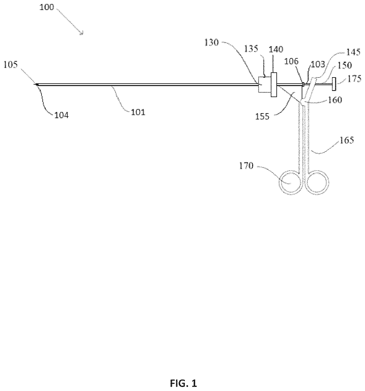
[0041]The following detailed description is merely exemplary in nature and is not intended to limit the described embodiments or the application and uses of the described embodiments. As used herein, the word “exemplary” or “illustrative” means “serving as an example, instance, or illustration.” Any implementation described herein as “exemplary” or “illustrative” is not necessarily to be construed as preferred or advantageous over other implementations. All of the implementations described below are exemplary implementations provided to enable persons skilled in the art to make or use the embodiments of the disclosure and are not intended to limit the scope of the disclosure, which is defined by the claims. For purposes of description herein, the terms “upper”, “lower”. “left”, “rear”, “right”. “front”, “vertical”, “horizontal”, and derivatives thereof shall relate to the invention as oriented in FIG. 1. Furthermore, there is no intention to be bound by any expressed or implied theo...


PUM


Abstract

The present invention is directed to an assembly used in minimally invasive procedures whereby laparoscopic instrument heads, prepositioned in the abdomen of a patient are securely doubly attached and detached internally while under direct laparoscopic visualization. The present invention permits surgeons to utilize full sized laparoscopic instrument heads on needlescopic instrument shafts. Multiple locking structures are employed to securely attach the laparoscopic instrument heads to a needlescopic instrument.

Description

CROSS REFERENCE TO RELATED APPLICATIONS[0001]This application claims priority from provisional U.S. Application No. 62 / 093,789 filed on Dec. 18, 2014 and entitled Method and Apparatus for Securing Laparoscopic Instrument Heads in the Abdomen under Direct Visualization.FIELD OF INVENTION[0002]The present application is directed toward an assembly used for remotely doubly attaching and detaching laparoscopic instrument heads within the abdomen of a patient to needlescopic instrument shafts under direct visualization.BACKGROUND OF THE INVENTION[0003]Laparoscopy is a surgical procedure performed through small incisions in the abdomen into which a trocar / cannula device is inserted to provide means for passage of medical instruments. A thin cylindrical instrument called a laparoscope, connected to a camera, is then used to provide a clear picture of the abdominal cavity to the surgeon.[0004]Prior to starting a surgical procedure a small incision is created through the abdominal wall of th...

Claims


Application Information

Patent Timeline
Patent Type & Authority Patents(United States)
IPC IPC(8): A61B17/00
CPCA61B17/00234A61B2017/00345A61B2017/00238A61B2017/0042A61B2017/00367A61B2017/00477A61B17/29A61B17/3417A61B2017/00464A61B2017/00473A61B2017/2931A61B2017/294A61B2017/3458
Inventor GOMEZ, RICARDO ALEXANDERHECK, SANDY LAWRENCECONLEY, ERIC WILLIAM
Owner NEW WAVE ENDO-SURGICAL CORP
Why Eureka
Try Eureka