Method and apparatus for generating quantitative data for biliary tree structures

a biliary and bile structure technology, applied in the field of quantitative data generation of biliary tree structures, can solve the problems of difficult detection using conventional mrcp and ercp techniques, occlusion related problems, and lack of depth information in mip visualization, so as to accurately and reliably assess the pathology of the patient's bile tree, accurate assessment of the bile tree, accurate and reliable assessment of topological and structural features

Active Publication Date: 2020-11-24
PERSPECTUM LTD
View PDF4 Cites 0 Cited by
  • Summary
  • Abstract
  • Description
  • Claims
  • Application Information

AI Technical Summary

Benefits of technology

[0012]Advantageously, by generating such quantitative biliary tree data comprising the generated quantitative structural parameters from volumetric medical imaging scan data, a clinician may be provided with enhanced information relating to the biliary tree of a patient that enables the clinician to more accurately and reliably assess the pathology of the patient's biliary tree, and in particular to more accurately and reliably assess topological and structural features of the biliary tree such as the presence of strictures and dilatations. In this manner, embodiments of the present invention provide an improved non-invasive diagnostic technique that enables a clinician to detect liver diseases, and more generally an improved non-invasive diagnostic technique for providing a clinician with an accurate assessment of the biliary tree.

Problems solved by technology

If either of the MRCP or ERCP procedures indicates the liver is normal (i.e. no Large Duct PSC detected), because of the cholestatic biochemical profile indicating the possible presence of PSC within the liver, the AASLD guidelines recommend a liver biopsy for diagnosing Small Duct PSC, which is difficult to detect using conventional MRCP and ERCP techniques.
MIP visualization suffers from a lack of depth information and occlusion related problems.
However, clinicians may be reluctant to proceed with an ERCP in the assessment of cholestasis due to its invasive nature and the use of x-radiation, and so PSC has likely been an underdiagnosed condition [“EASL clinical practice guidelines”, European Association for the Study of the Liver, Hepatology 2009; 51:237-267].

Method used


Image

  • Method and apparatus for generating quantitative data for biliary tree structures
  • Method and apparatus for generating quantitative data for biliary tree structures
  • Method and apparatus for generating quantitative data for biliary tree structures

Examples


Embodiment Construction

[0061]The present invention will now be described with reference to the accompanying drawings in which there is illustrated an example of a method and apparatus for generating quantitative data for biliary tree structures from volumetric medical imaging scan data. However, it will be appreciated that the present invention is not limited to the specific examples herein described and as illustrated in the accompanying drawings.

[0062]Furthermore, because the illustrated embodiments of the present invention may for the most part, be implemented using electronic components and circuits known to those skilled in the art, details will not be explained in any greater detail than that considered necessary as illustrated below, for the understanding and appreciation of the underlying concepts of the present invention and in order not to obfuscate or distract from the teachings of the present invention.

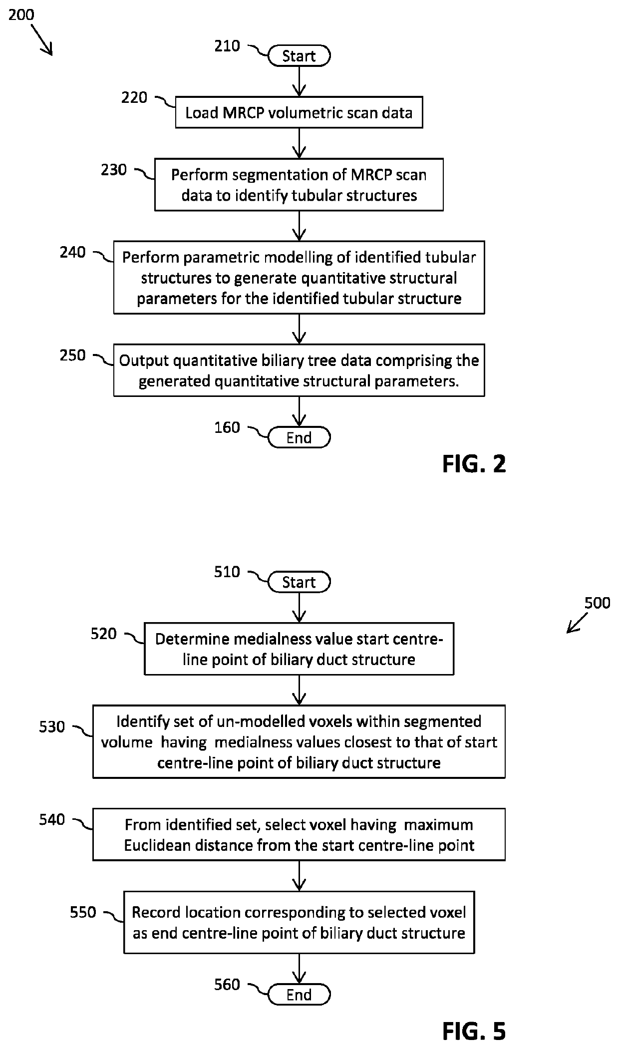
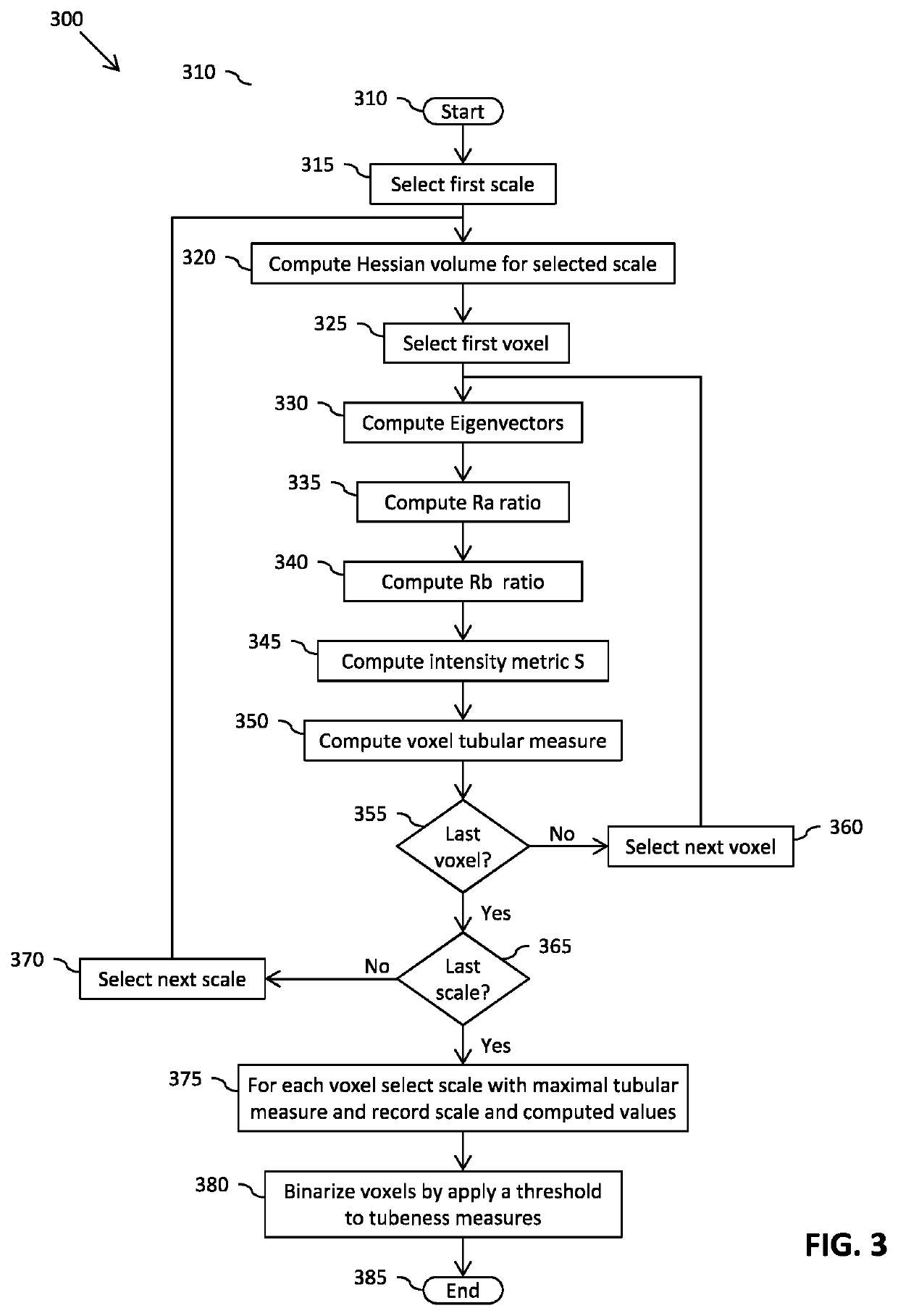
[0063]Referring now to FIG. 2, there is illustrated a simplified flowchart 200 of an example...


PUM


Abstract

A method (200) and apparatus (1100) for generating quantitative data for biliary tree structures from volumetric medical imaging scan data. The method comprises performing segmentation (230; 300) of a volume of the medical imaging scan data to identify tubular biliary structures within the volume of the medical imaging scan data; for at least one segmented tubular biliary structure within the volume of the medical imaging scan data, computing (240; 800) at least one set of quantitative structural parameters for at least one location along the length of the tubular biliary structure; and outputting (250) quantitative biliary tree data comprising the at least one set of quantitative structural parameters for the at least one segmented tubular biliary structure.

Description

FIELD OF THE INVENTION[0001]This invention relates to a method and apparatus for generating quantitative data for biliary tree structures. In particular to a method and apparatus for generating quantitative data for biliary tree structures from volumetric medical imaging scan data.BACKGROUND OF THE INVENTION[0002]For the diagnosis of certain liver diseases, it is often critical for the diagnosing clinician to obtain an accurate assessment of the biliary tree. For example, FIG. 1 illustrates the recommendations for diagnosing Primary Sclerosing Cholangitis (PSC) as documented in the American Association for the Study of Liver Diseases (AASLD) practice guidelines.[0003]Upon a cholestatic biochemical profile indicating the possible presence of PSC within a patient's liver, the AASLD guidelines recommend an ultrasound imaging scan be performed that can enable the clinician to observe biliary duct wall thickening and / or focal biliary duct dilatations.[0004]If the ultrasound scan is non-d...

Claims


Application Information

Patent Timeline
Patent Type & Authority Patents(United States)
IPC IPC(8): G06K9/00G06T7/12G06T7/162G06T7/62A61B5/055A61B5/00G06T7/00
CPCG06T7/62A61B5/055G06T7/0012G06T7/12G06T7/162A61B5/4244G06T2207/30172G06T2200/04G06T2207/30056A61B2576/02G06T2207/10088G06T7/10G06T2207/10064G06T2207/10068
Inventor VIKAL, SIDDHARTHBRADY, JOHN MICHAEL
Owner PERSPECTUM LTD
Why Eureka
Try Eureka