Microporous hydrogel scaffolds for cell transplantation

a micro-porous hydrogel and cell technology, applied in the field of biomaterial implants and methods, can solve the problems of preventing neovascularization and hindering the mass transport of nutrients to encapsulated cells, and achieve the effects of facilitating blood glucose sensing, preventing neovascularization and hindering the mass transport of nutrients

Active Publication Date: 2021-04-13
RGT UNIV OF MICHIGAN
View PDF11 Cites 0 Cited by
  • Summary
  • Abstract
  • Description
  • Claims
  • Application Information

AI Technical Summary

Benefits of technology

[0005]The present disclosure is generally directed to islet cell transplantation onto microporous scaffolds that allow for revascularization of the transplanted islets. The ability to revascularize the islets distinguishes this approach from encapsulation systems, as revascularization can provide the nutrients necessary for islet survival while also facilitating the sensing of blood glucose and the distribution of insulin. Islet encapsulation technologies using hydrogels for extrahepatic islet transplantation have become widespread in the cell transplantation field, due to their ability to shield encapsulated cells from the host's immune system. However, previous technologies are limited by preventing neovascularization and hindering mass transport of nutrients to encapsulated cells.
[0007]In various embodiments of the present disclosure, these microporous scaffolds have been functionalized to control the local environment. In at least some embodiments, the microporous scaffolds are functionalized (e.g., with extracellular matrix proteins and / or trophic factors) to enhance the engraftment of the transplanted islets. Additionally or alternatively, in some embodiments, the microporous scaffolds are functionalized (e.g., with immune cytokines and / or Tregs) to modulate the innate and adaptive immune responses to delay rejection or promote immune tolerance. Accordingly, in various embodiments, the microporous scaffolds have been functionalized to increase success of cell transplantation.

Problems solved by technology

However, previous technologies are limited by preventing neovascularization and hindering mass transport of nutrients to encapsulated cells.

Method used


Image

  • Microporous hydrogel scaffolds for cell transplantation
  • Microporous hydrogel scaffolds for cell transplantation
  • Microporous hydrogel scaffolds for cell transplantation

Examples


example 1

Microporous Scaffolds Support Engraftment of Mouse and Human Islets

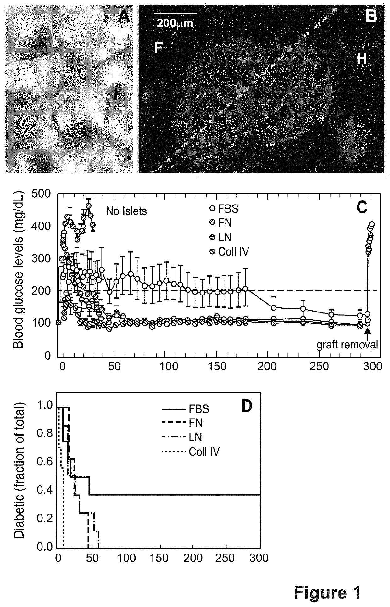
[0080]Microporous scaffolds for transplantation of islets into epididymal fat pads have been developed, which are clinically equivalent to the omentum [Blomeier et al., Transplantation. 2006; 82:452-9.]. Mouse islets seeded onto the microporous scaffolds quickly distributed within the scaffold pores, with 1-2 islets per pore (FIG. 1A). Fourteen days after implantation into a syngeneic host, islets were seen to maintain their morphology, demonstrate a dense and functional vasculature (staining for tomato lectin) (FIG. 1B), and additionally contain proliferating cells as indicated by PCNA staining, suggesting that turnover and remodeling were occurring within the islets (data not shown) [Gibly et al., Biomaterials. 2011; 32:9677-84]. The engrafted islets reversed host diabetes for 300 days (FIG. 1C), and hyperglycemia returned after explant of the fat pad (FIG. 1C). Interestingly, the scaffold lost integrity around 100...

example 2

[0085]In some aspects, the present disclosure is directed toward the use of poly(ethylene glycol) (PEG) microporous scaffolds as a safe, efficient, and clinically applicable approach for transplantation of islets or hESC-derived n-cell progenitors. Questions examined in the examples relate to the scaffold design needed to create a supportive environment that maximally promotes engraftment and function of transplanted islets, or maturation of hESC-derived n-cell progenitors and subsequent normalization of blood glucose levels. Furthermore, the scaffolds are non-degradable which allows the implant to be explanted should the need arise; accordingly, these studies also assessed the efficacy of explantation at recovering transplanted cells.

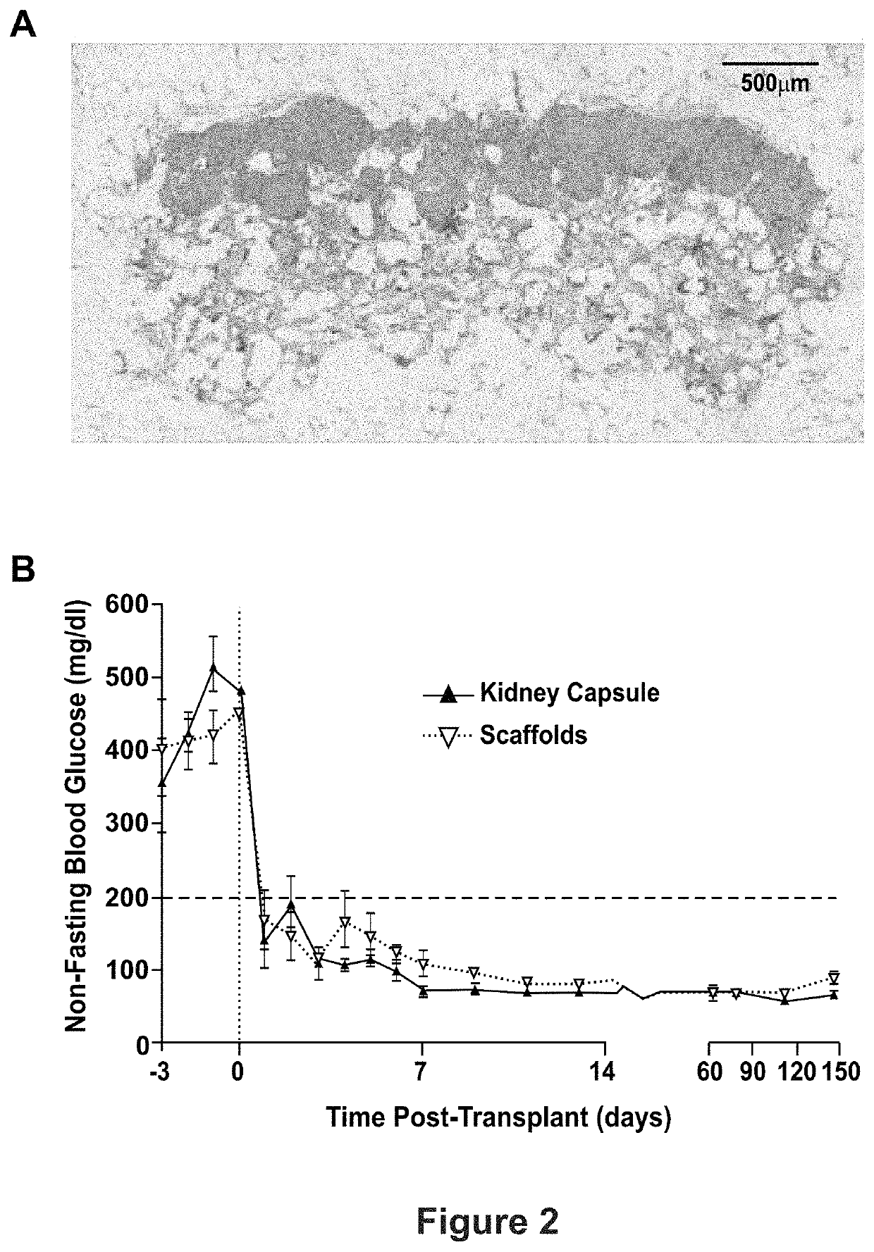
[0086]Research over the past 10 years with microporous scaffolds has demonstrated that transplanted islets engraft, become revascularized, and function to maintain normal blood glucose levels for long times (FIG. 1C). These scaffolds have supported the...

example 3

Investigate Transplantation of Allogeneic Islets into the Peritoneal Fat on Microporous PEG Scaffolds for Long-Term Graft Function and Assess the Ability for Complete Graft Retrieval

[0087]In this example, results with microporous scaffolds are extended to investigate the potential for complete graft retrieval in rodent models, as well as the transplantation and retrieval of allogeneic islets delivered on scaffolds using a non-human primate model. Prior studies using microporous scaffolds composed of poly(lactic-co-glycolide) (PLG) or PEG indicate that this platform supports the efficient transplantation, engraftment, and function of transplanted islets. Both material platforms have had similar efficacy in islet transplantation, and can be functionalized (i.e., ECM proteins, trophic factors) in analogous manners (described below and Example 4). PEG-based microporous scaffolds are investigated as the material is expected to have a decreased inflammatory response relative to PLG, have ...


PUM

PropertyMeasurementUnit
pore sizeaaaaaaaaaa
pore sizeaaaaaaaaaa
diameteraaaaaaaaaa

Abstract

The present disclosure relates generally to biomaterial implants and methods for delivering a cell to an individual in need thereof and, more particularly, to biomaterial implants and techniques for delivering islets and / or β-cell progenitors to an individual.

Description

CROSS-REFERENCE TO RELATED APPLICATION[0001]This application claims the priority benefit under 35 U.S.C. § 119(e) of Provisional U.S. Patent Application No. 62 / 442,768, filed Jan. 5, 2017, the disclosure of which is incorporated herein by reference in its entirety.FIELD OF THE DISCLOSURE[0002]The present disclosure relates generally to biomaterial implants and methods for delivering a cell to an individual in need thereof and, more particularly, to biomaterial implants and techniques for delivering an islet cell and / or a β-cell progenitor to an individual.BACKGROUND[0003]The background description provided herein is for the purpose of generally presenting the context of the disclosure. Work of the presently named inventor, to the extent it is described in this background section, as well as aspects of the description that may not otherwise qualify as prior art at the time of filing, are neither expressly nor impliedly admitted as prior art against the present disclosure.[0004]Clinic...

Claims


Application Information

Patent Timeline
Patent Type & Authority Patents(United States)
IPC IPC(8): A61L27/56A61L27/18A61L27/24A61L27/54A61L27/38A61L27/22A61L27/36
CPCA61L27/56A61L27/18A61L27/222A61L27/24A61L27/3633A61L27/38A61L27/3804A61L27/54A61L2300/414A61L2300/43C08L71/02
Inventor KASPUTIS, TADASSKOUMAL, MICHAELSHEA, LONNIE D.
Owner RGT UNIV OF MICHIGAN
Why Eureka
Try Eureka