
Generating synthesized projection images for 3D breast tomosynthesis or multi-mode x-ray breast imaging

a technology of breast imaging and synthesizing, which is applied in the direction of image enhancement, patient positioning for diagnostics, instruments, etc., can solve the problem of increasing the total amount of time required to complete the imaging process

Active Publication Date: 2021-08-17
HOLOGIC INC
View PDF426 Cites 3 Cited by
  • Summary
  • Abstract
  • Description
  • Claims
  • Application Information

AI Technical Summary

Benefits of technology

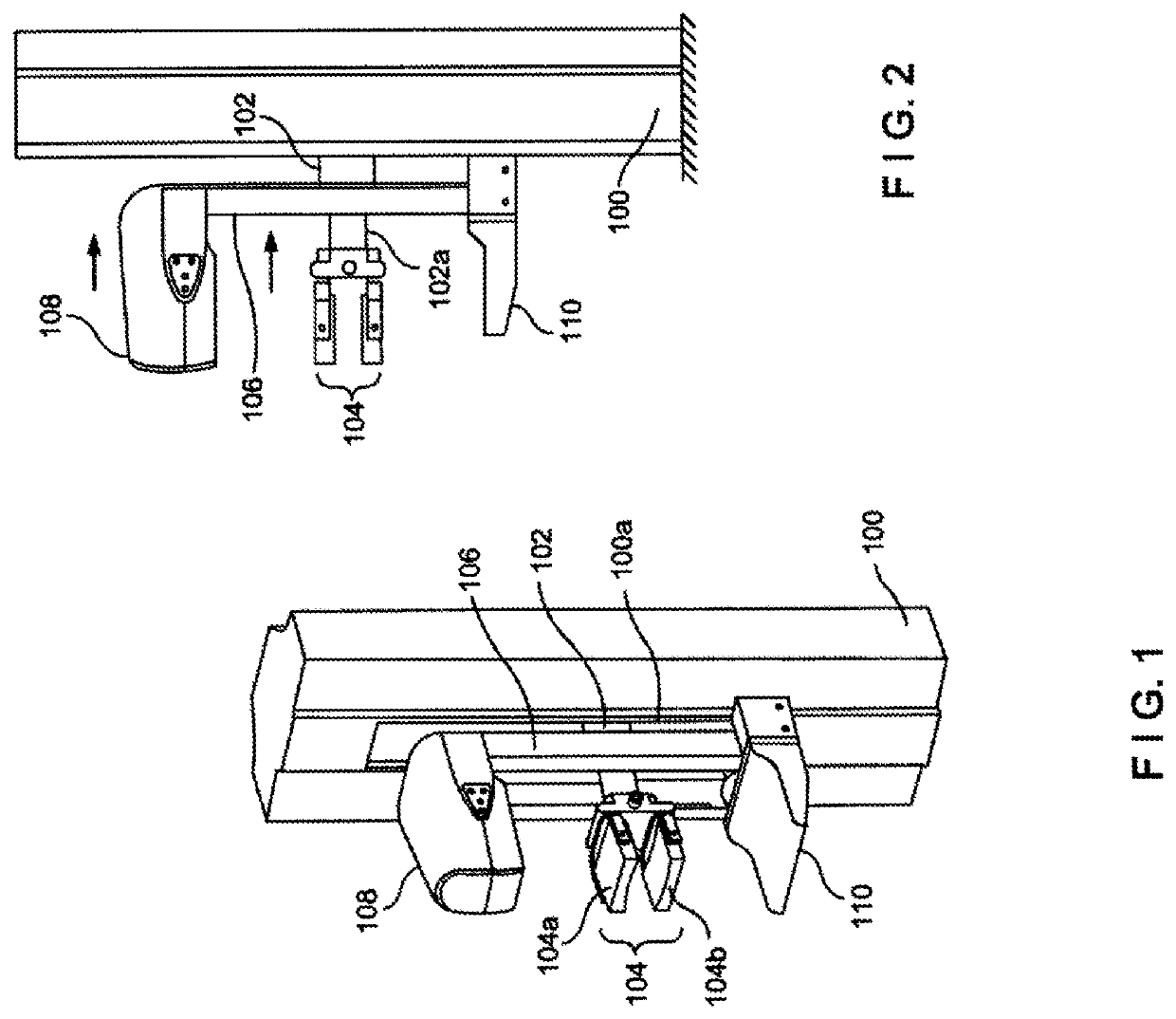
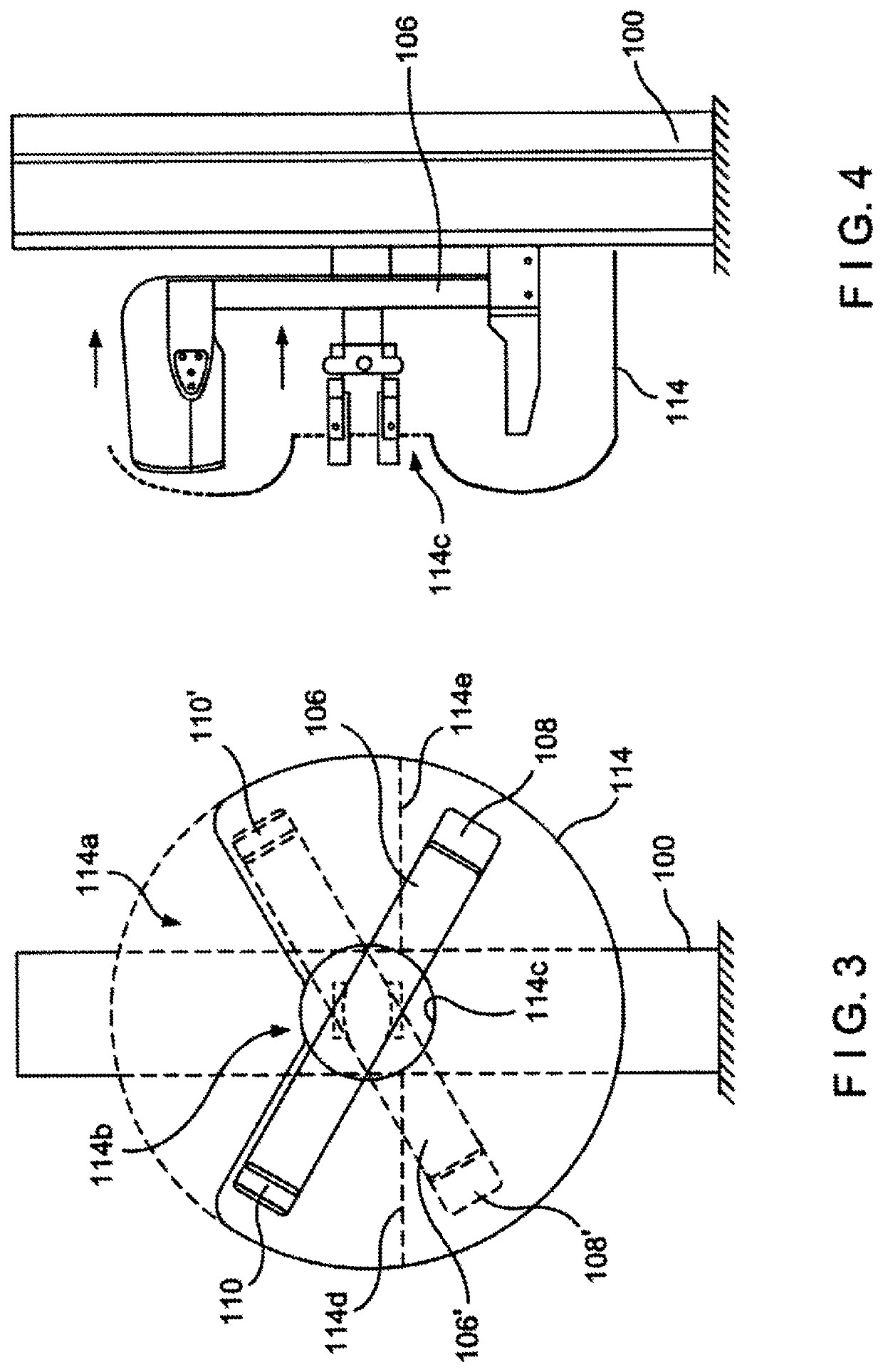
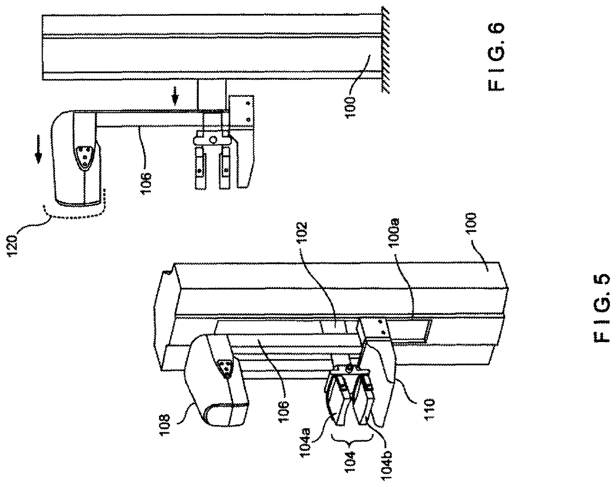
[0004]Examples of the present disclosure describe systems and methods for medical imaging through the use of a synthesized virtual projections generated from real projections. In an aspect, the technology relates to a system for generating images of a breast. The system includes an x-ray source, an x-ray detector, at least one processor operatively connected to the x-ray detector, and memory operatively connected to the at least one processor, the memory storing instructions that, when executed by the at least one processor, cause the system to perform a set of operations. The operations include, emitting, from the x-ray source, a first x-ray emission at a first angular location relative to the x-ray detector; detecting, by the x-ray detector, the first x-ray emission after passing through the breast; generating first x-ray imaging data from the detected first x-ray emission; emitting, from the x-ray source, a second x-ray emission at a second angular location relative to the breast; detecting, by the x-ray detector, the second x-ray emission after passing through the breast; generating second x-ray imaging data from the detected second x-ray emission; synthesizing, based on at least the first x-ray imaging data and the second x-ray imaging data, third x-ray imaging data for a third angular location relative to the breast, wherein the third angular location is different from the first angular location and the second angular location, thereby eliminating the need for an x-ray emission at the third angular location; and generating and displaying an image of the breast from the third x-ray imaging data.

Problems solved by technology

Acquiring each projection image, however, increases the total amount of time required to complete the imaging process.

Method used


Image

  • Generating synthesized projection images for 3D breast tomosynthesis or multi-mode x-ray breast imaging
  • Generating synthesized projection images for 3D breast tomosynthesis or multi-mode x-ray breast imaging
  • Generating synthesized projection images for 3D breast tomosynthesis or multi-mode x-ray breast imaging

Examples


Embodiment Construction

[0029]As discussed above, a tomosynthesis system acquires a series of x-ray projection images, each projection image obtained at a different angular displacement as the x-ray source moves along a path, such as a circular arc, over the breast. More specifically, the technology typically involves taking two-dimensional (2D) real projection images of the immobilized breast at each of a number of angles of the x-ray beam relative to the breast. The resulting x-ray measurements are computer-processed to reconstruct images of breast slices that typically are in planes transverse to the x-ray beam axis, such as parallel to the image plane of a mammogram of the same breast, but can be at any other orientation and can represent breast slices of selected thicknesses. Acquiring each real projection image introduces additional radiation to the patient and increases the total amount of time required to complete the imaging process. The use of fewer real projection images, however, leads to worse...


PUM


Abstract

Methods and systems for medical imaging including synthesizing virtual projections from acquired real projections and generating reconstruction models and images based on the synthesized virtual projections and acquired real projections. For example, first x-ray imaging data is generated from a detected first x-ray emission at a first angular location and second x-ray imaging data is generated from a detected second x-ray emission at a second angular location. Based on at least the first x-ray imaging data and the second x-ray imaging data, third x-ray imaging data for a third angular location relative to the breast may be synthesized. An image of the breast may be displayed or generated from the third x-ray imaging data.

Description

RELATED CASES[0001]This application claims the benefit of U.S. Provisional Application No. 62 / 730,818 filed on Sep. 13, 2018, which is hereby incorporated by reference in its entirety.BACKGROUND[0002]Medical imaging has become a widely used tool for identifying and diagnosing abnormalities, such as cancers or other conditions, within the human body. Medical imaging processes such as mammography and tomography are particularly useful tools for imaging breasts to screen for, or diagnose, cancer or other lesions with the breasts. Tomosynthesis systems are mammography systems that allow high resolution breast imaging based on limited angle tomosynthesis. Tomosynthesis, generally, produces a plurality of x-ray images, each of discrete layers or slices of the breast, through the entire thickness thereof. In contrast to typical two-dimensional (2D) mammography systems, a tomosynthesis system acquires a series of x-ray projection images, each projection image obtained at a different angular...

Claims


Application Information

Patent Timeline
Patent Type & Authority Patents(United States)
IPC IPC(8): A61B6/00A61B6/04G06T11/00G06T7/70G06T7/00A61B5/00A61B6/02G06T5/50
CPCA61B6/502A61B5/7267A61B6/025A61B6/4028A61B6/5235G06T5/50G06T7/0012G06T7/70G06T11/003G06T2207/10081G06T2207/10112G06T2207/20081G06T2207/20084G06T2207/20221G06T2207/30068G06T2207/30168A61B6/032A61B6/54G06T11/006
Inventor ZHANG, YIHENGJING, ZHENXUERUTH, CHRISTOPHER
Owner HOLOGIC INC
Learn More