Cardiac computed tomography

a computed tomography and cardiac technology, applied in tomography, diagnostic recording/measuring, applications, etc., can solve problems such as dose increas

Active Publication Date: 2022-05-10
KONINKLJIJKE PHILIPS NV
View PDF4 Cites 0 Cited by
  • Summary
  • Abstract
  • Description
  • Claims
  • Application Information

AI Technical Summary

Benefits of technology

The patent aims to reduce the amount of radiation used in cardiac computed tomography (CT) while improving image quality. This is particularly useful for procedures such as coronary CT and coronary CT angiography.

Problems solved by technology

This may allow an adjustment of the projection angle ranges used for image reconstruction, but also results also results in increase of dose.

Method used


Image

  • Cardiac computed tomography
  • Cardiac computed tomography

Examples


Embodiment Construction

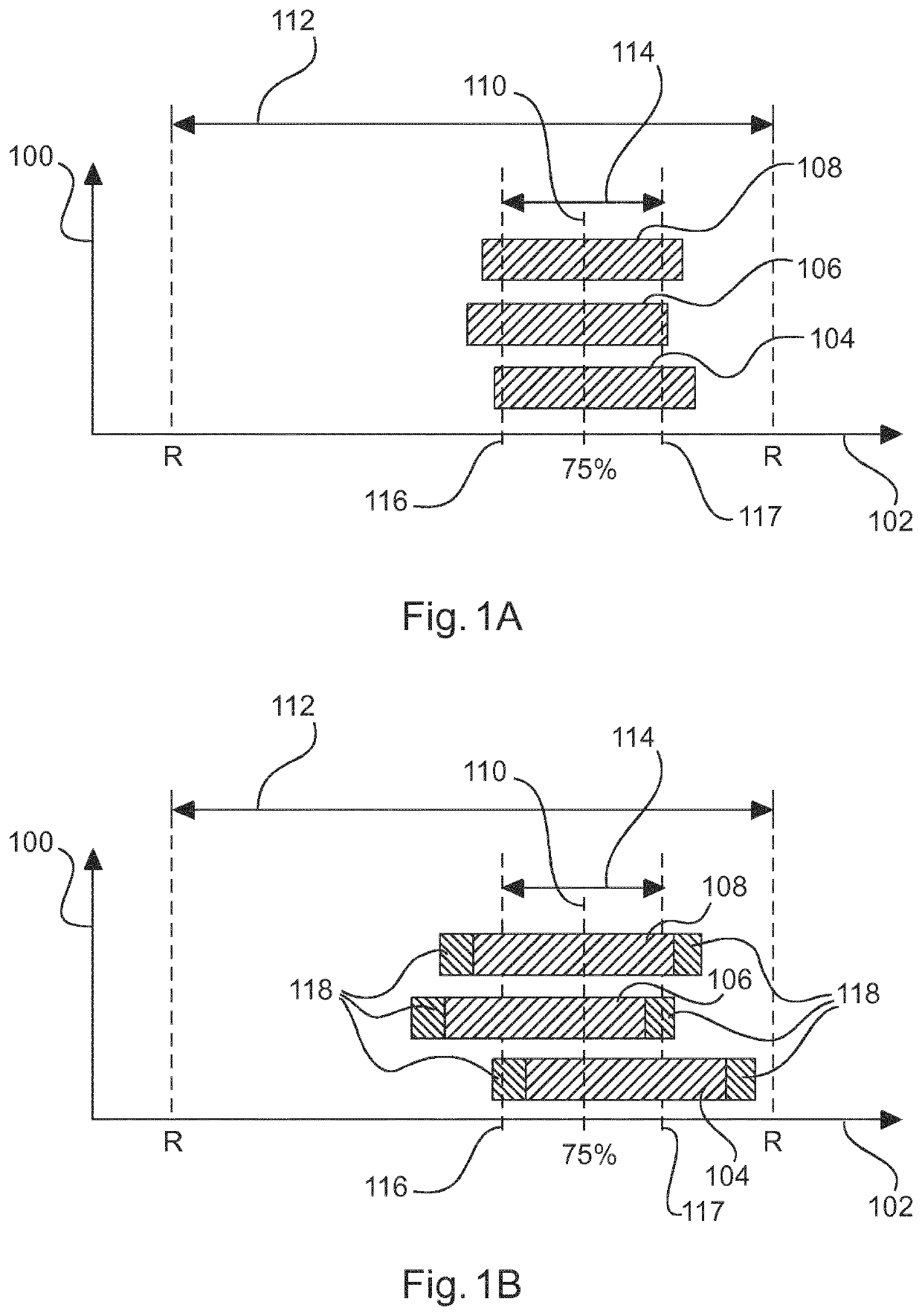
[0046]FIGS. 1A and 1B each schematically show acquisition time periods as a function of the heart phase φ. FIGS. 1A and 1B thereby illustrate findings and insights, on which the present invention is based. More specifically, the y-axis 100 in FIGS. 1A and 1B, respectively, depicts a number of shoots, i.e., acquisition time intervals and / or acquisition time periods, of a step-and-shoot scan performed with a cardiac CT imaging system, and the x-axis 102 depicts the heart phase φ. Therein, a total number of three shots 104, 106, 108 of the step-and-shoot scan are shown.

[0047]Generally, prospective electrocardiography-triggering (ECG-triggering) of the CT imaging system has been applied during acquisition of projection data, which was started at a certain time after an R-peak of a heart cycle 112 was determined based on ECG data and / or based on a dataset comprising ECG data. Therein, the heart cycle 112 is determined as time period between two consecutive R-peaks in the ECG data.

[0048]I...


PUM


Abstract

A method for operating a cardiac CT imaging system (200) in coronary CT with prospective electrocardiography-triggering of the imaging system (200) is proposed. The method comprises the steps of determining a plurality of R-peak times (ti) from a dataset of electrocardiography data, determining a variability of a heart rate by statistically analyzing the determined R-peak times (ti), and automatically adjusting an acquisition time period (ΔTac) of the CT imaging system (200) for acquiring projection data based on the determined variability of the heart rate.

Description

CROSS REFERENCE TO RELATED APPLICATIONS[0001]This application is the U.S. National Phase application under 35 U.S.C. § 371 of International Application No. PCT / EP2017 / 072038 filed Sep. 4, 2017, published as WO 2018 / 046413 on Mar. 15, 2018, which claims the benefit of European Patent Application Number 16187741.0 filed Sep. 8, 2016. These applications are hereby incorporated by reference herein.FIELD OF THE INVENTION[0002]The invention generally relates to cardiac computed tomography (CT). More specifically, the invention relates to a method for operating a cardiac CT imaging system, to a cardiac CT imaging system, to a computer program element and to a computer-readable medium.BACKGROUND OF THE INVENTION[0003]Modern computed tomography imaging systems utilized for imaging a heart, e.g. for coronary CT and / or for coronary CT angiography, are usually based on electrocardiography (ECG) data for determining an acquisition time and / or an acquisition time period, in which projection data ...

Claims


Application Information

Patent Timeline
IPC IPC(8): A61B6/00A61B6/03A61B5/352A61B5/024G06F17/18
CPCA61B6/541A61B5/02405A61B5/352A61B6/032A61B6/503G06F17/18A61B6/5205
Inventor THRAN, AXEL
Owner KONINKLJIJKE PHILIPS NV
Why Eureka
Try Eureka