Therapeutic drug for diseases related to endoplasmic reticulum cell death in corneal endothelium
a technology of endoplasmic reticulum cells and therapeutic drugs, which is applied in the direction of antibody medical ingredients, peptide/protein ingredients, and metabolic disorders, etc., can solve the problem that human corneal endothelial cells have very limited regeneration ability
- Summary
- Abstract
- Description
- Claims
- Application Information
AI Technical Summary
Benefits of technology
Problems solved by technology
Method used
Image

Examples
preparation example 1
ized Corneal Endothelial Cell Line (iFECD) Model from Fuchs' Endothelial Corneal Dystrophy Patient
[0115]In the present example, an immobilized corneal endothelial cell line (iFECD) was made from corneal endothelial cells from Fuchs' endothelial corneal dystrophy patients.
(Culture Method)
[0116]Corneal endothelial cells were mechanically peeled off with a basal membrane from a corneal for research purchased from the Seattle Eye Bank. After collagenase was used to detach and collect the corneal endothelial cell from the basal membrane, the cells were subjected to primary culture. For a medium, Opti-MEM I Reduced-Serum Medium, Liquid (INVITROGEN catalog No.: 31985-070) to which 8% FBS (BIOWEST, catalog No.: S1820-500), 200 mg / ml of CaCl2.2H2O (SIGMA catalog No.: C7902-500G), 0.08% of chondroitin sulfate (SIGMA catalog No.: C9819-5G), 20 μg / ml of ascorbic acid (SIGMA catalog No.: A4544-25G), 50 μg / ml of gentamicin (INVITROGEN catalog No.: 15710-064) and 5 ng / ml of EGF (INVITROGEN catalog...
preparation example 2
l Functioning of Immobilized Corneal Endothelial Cell Line (iFECD)
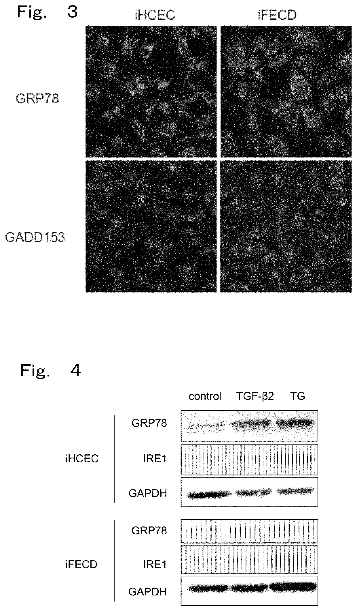
[0119]Normal functioning of an immobilized corneal endothelial cell line (iFECD) was confirmed in the present Example.
(Immunostaining by Na+ / K+-ATPase and ZO-1)
[0120]First, immunostaining was performed with Na+ / K+-ATPase and ZO-1 to confirm normal functioning of an immobilized corneal endothelial cell line (iFECD). This is for examining the functions of corneal endothelial cells, i.e., pumping and barrier functions. Na+ / K+-ATPase and ZO-1 indicates normal pumping and barrier functions of corneal endothelial cells, respectively. The technology is as follows.
[0121](Method of Observing Cells with Staining or the Like (Histological Test))
[0122]Cells were observed with a phase difference microscope. After cells were immobilized, immunostaining was applied with ZO-1 and Na+ / K+-ATPase as function associated markers for observation with a fluorescent microscope. For a tissue staining test, cultured cells were placed in Lab-Te...
example 1
ical Observation of Endoplasmic Reticulum and Mitochondria in Fuchs' Endothelial Corneal Dystrophy
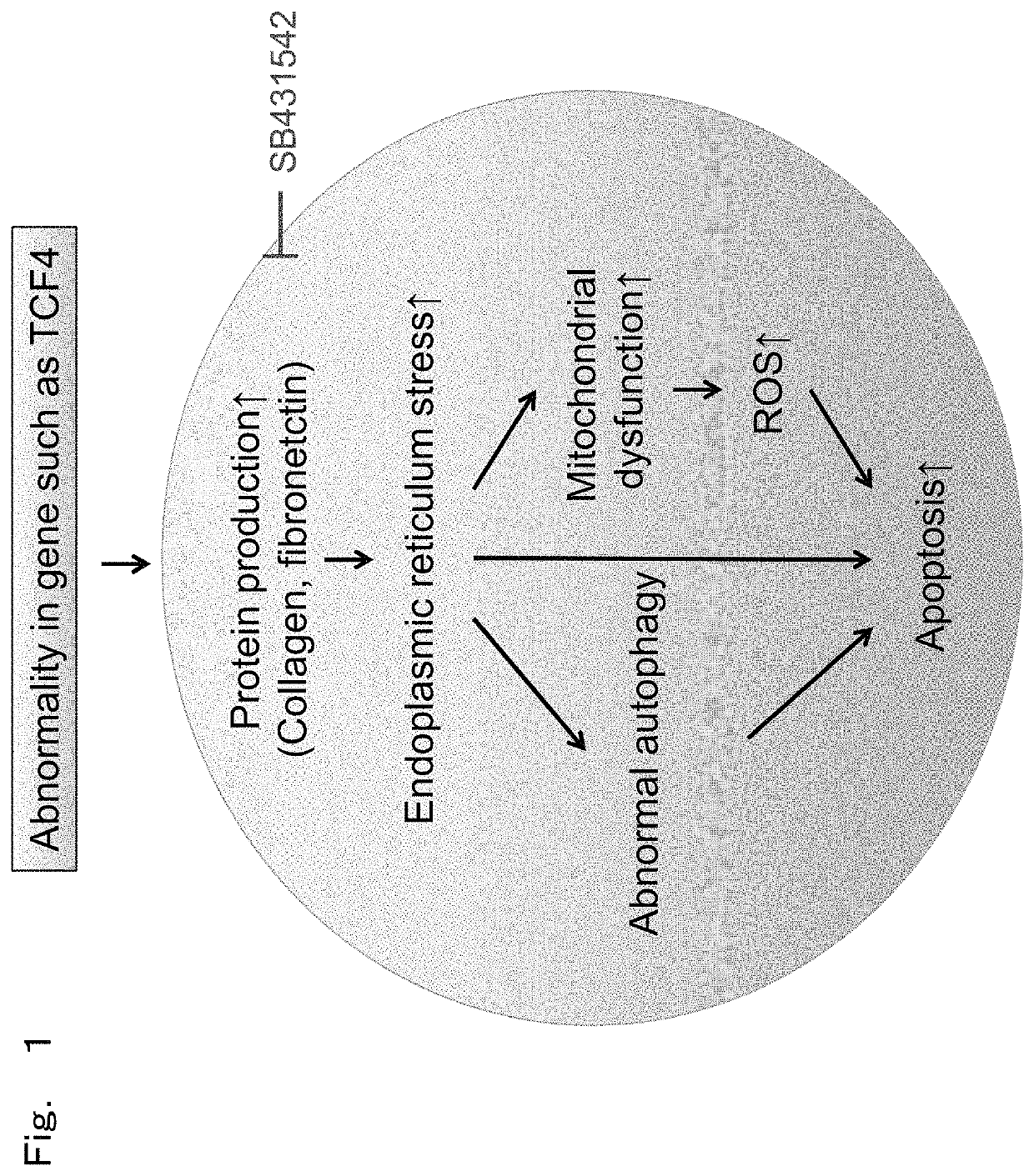
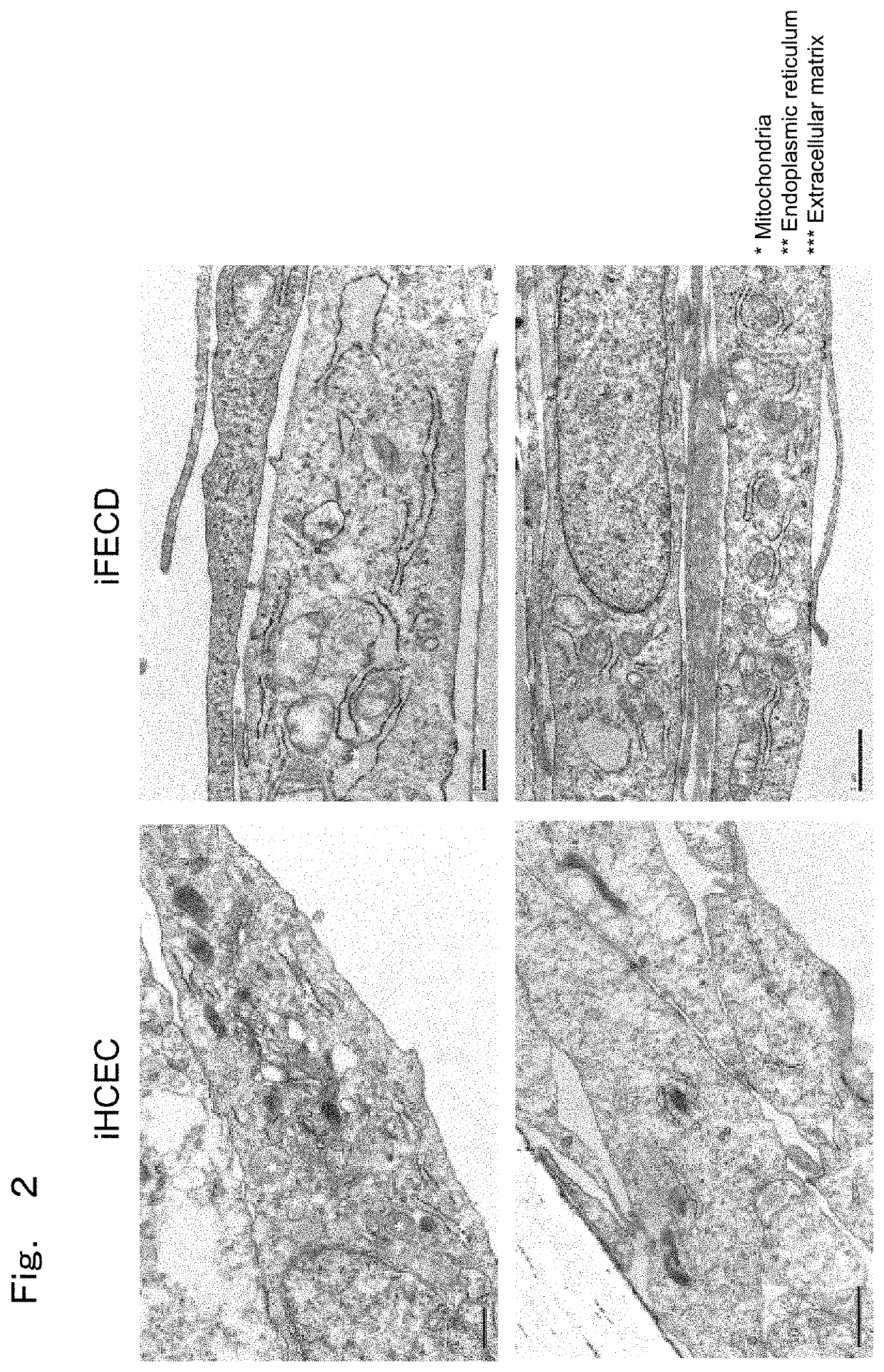
[0126]The present Example observed whether morphological abnormality is observed in endoplasmic reticulum and mitochondria in cells with Fuchs' endothelial corneal dystrophy model prepared in the above-described Preparation Example as a representative example in order to study endoplasmic reticulum stress.
(Observation of Organelles by Electron Microscope)
[0127]Cells were then observed with an electron microscope to confirm elevation in ECM production and ER stress. As a pre-immobilization, a transwell seeded with cells was immobilized for three hours at room temperature with 2.5% glutaraldehyde with a pH 7.2 diluted with 0.1 M sodium cacodylate and washed three times with 0.1 M sodium cacodylate. Then, as post immobilization, it was immobilized for 1 hour at room temperature with 1% osmic acid diluted with 0.1 M sodium cacodylate and washed three times with distilled water. In order to ...
PUM
| Property | Measurement | Unit |
|---|---|---|
| concentration | aaaaa | aaaaa |
| concentration | aaaaa | aaaaa |
| concentration | aaaaa | aaaaa |
Abstract
Description
Claims
Application Information
 - R&D Engineer
- R&D Manager
- IP Professional
- Industry Leading Data Capabilities
- Powerful AI technology
- Patent DNA Extraction
Browse by: Latest US Patents, China's latest patents, Technical Efficacy Thesaurus, Application Domain, Technology Topic.
© 2024 PatSnap. All rights reserved.Legal|Privacy policy|Modern Slavery Act Transparency Statement|Sitemap