Navigational guidance via computer-assisted fluoroscopic imaging
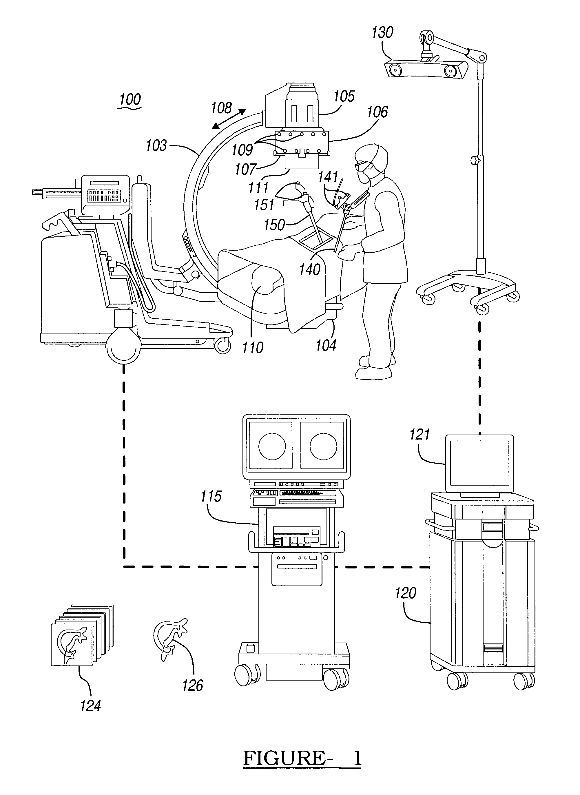
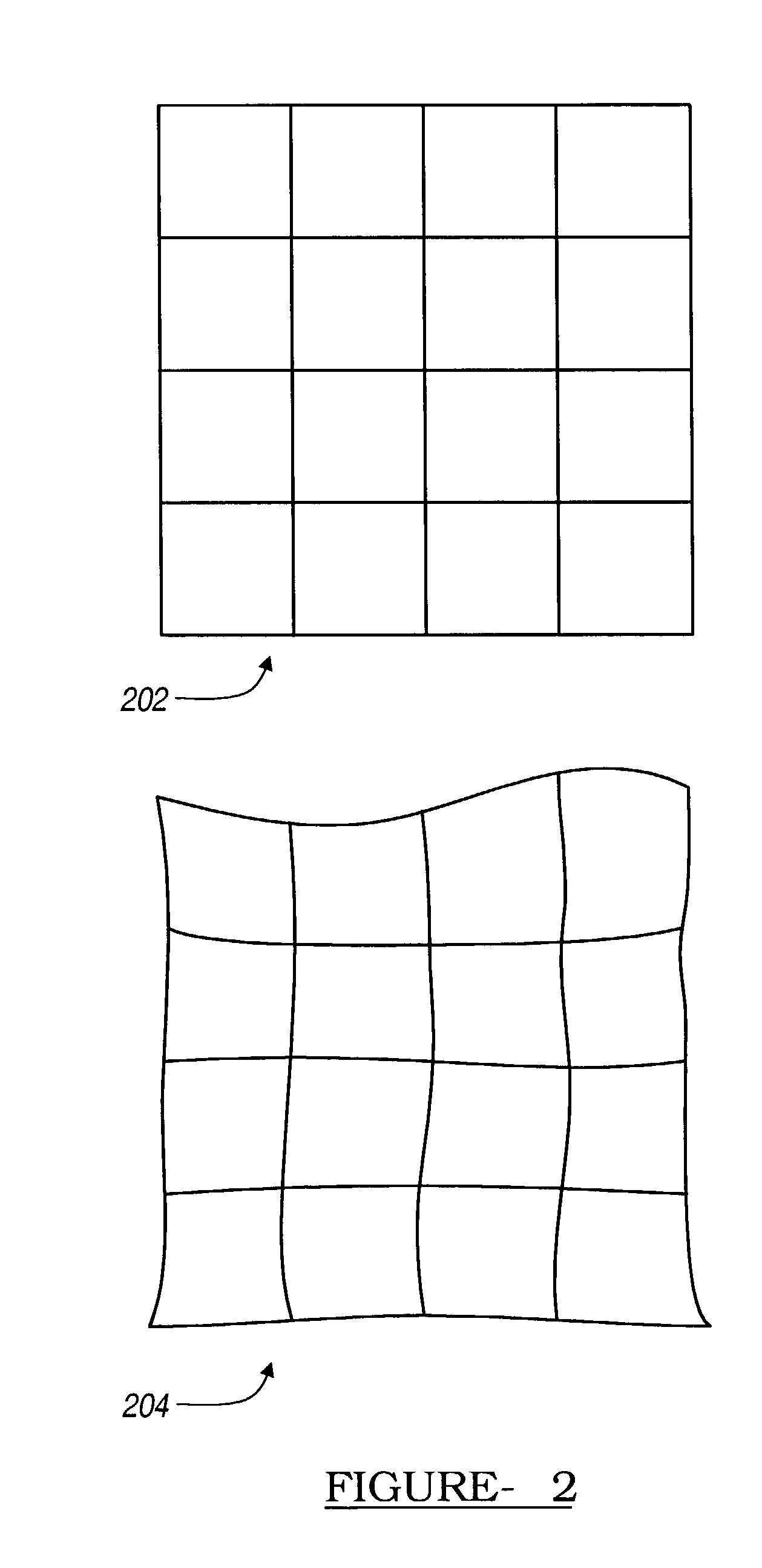
a technology of navigation guidance and computer assisted fluoroscopic imaging, which is applied in the direction of instruments, catheters, applications, etc., can solve the problems of inability to view, inability to accurately calculate the distance between the image and the receiving section, and the image generated by the receiving section 105 tends to suffer from undesirable distortion,
- Summary
- Abstract
- Description
- Claims
- Application Information
AI Technical Summary
Problems solved by technology
Method used
Image
Examples
example 2
Acquisition of an X-Ray View Down the Medial Axis of a Vertebral Pedicle
[0097] In certain clinical procedures, it may be desirable to acquire a fluoroscopic x-ray image view looking substantially straight down the medial axis of a vertebral pedicle. For the purposes of this example, a vertebral pedicle can be thought of as a cylinder, and the medial axis corresponds to the central axis of the cylinder.
[0098] FIG. 15A is an x-ray image in which the view direction of the imager is aligned with the medial axis of the pedicle (i.e., the medial axis of the pedicle is into the plane of the image). In this so-called "owl's eye" view, the pedicle appears as circle 1501 within the image. It is often difficult to precisely acquire this view using only fluoroscopic x-ray images, as it is difficult to align the view direction of imager 100 with the medial axis of the pedicle using only fluoroscopic images.
[0099] Given an anterior / posterior fluoroscopic image view of the spine, such as the one s...
example 3
Use of Digitally Reconstructed Radiography in the Placement of a Surgical Implant
[0101] With conventional fluoroscopic x-ray image acquisition, radiation passes through a physical media to create a projection image on a radiation sensitive film or an electronic image intensifier. Given a 3D CT data set, a simulated x-ray image can also be generated using a technique known as digitally reconstructed radiography (DRR). DRR is well known in the art, and is described, for example, by L. Lemieux et al., "A Patient-to-Computed-Tomography Image Registration Method Based on Digitally Reconstructed Radiographs," Medical Physics 21(11), pp 1749-1760, November 1994.
[0102] When a DRR image is created, a fluoroscopic image is formed by computationally projecting volume elements (voxels) of the 3D CT data set onto a selected image plane. Using a 3D CT data set of a given patient, it is possible to create a DRR image that appears very similar to a corresponding x-ray image of the same patient. A r...
example 4
Obtaining a Particular Two-Dimensional View Direction Using Digitally Reconstructed Radiograph Images
[0106] The DRR technique can be used to provide guidance to the physician when acquiring an owl's eye view of a vertebral pedicle. Given a three-dimensional CT data set containing a vertebra and associated pedicle, the physician may use computer 120 to manually locate a three-dimensional representation of the pedicle's medial axis relative to the three-dimensional images of the vertebrae. Once this placement has been achieved, it is possible to synthesize an owl's eye view of the vertebrae based upon the view direction specified by the physician's selection of the three-dimensional medial axis. This synthetic image can then be displayed to the surgeon during surgery and used to guide the acquisition of an actual owl's eye view using the fluoroscope. By visually comparing fluoroscopic images taken while positioning the fluoroscope to the synthetic owl's eye view, the physician can acq...
PUM
 Abstract
Description
Claims
Application Information
 - R&D
- Intellectual Property
- Life Sciences
- Materials
- Tech Scout
- Unparalleled Data Quality
- Higher Quality Content
- 60% Fewer Hallucinations
Browse by: Latest US Patents, China's latest patents, Technical Efficacy Thesaurus, Application Domain, Technology Topic, Popular Technical Reports.
© 2025 PatSnap. All rights reserved.Legal|Privacy policy|Modern Slavery Act Transparency Statement|Sitemap|About US| Contact US: help@patsnap.com


