
System and method for x-ray based assessment of aneurysm pulsation

Active Publication Date: 2009-03-12
SIEMENS MEDICAL SOLUTIONS USA INC
View PDF6 Cites 28 Cited by
  • Summary
  • Abstract
  • Description
  • Claims
  • Application Information

AI Technical Summary

Benefits of technology

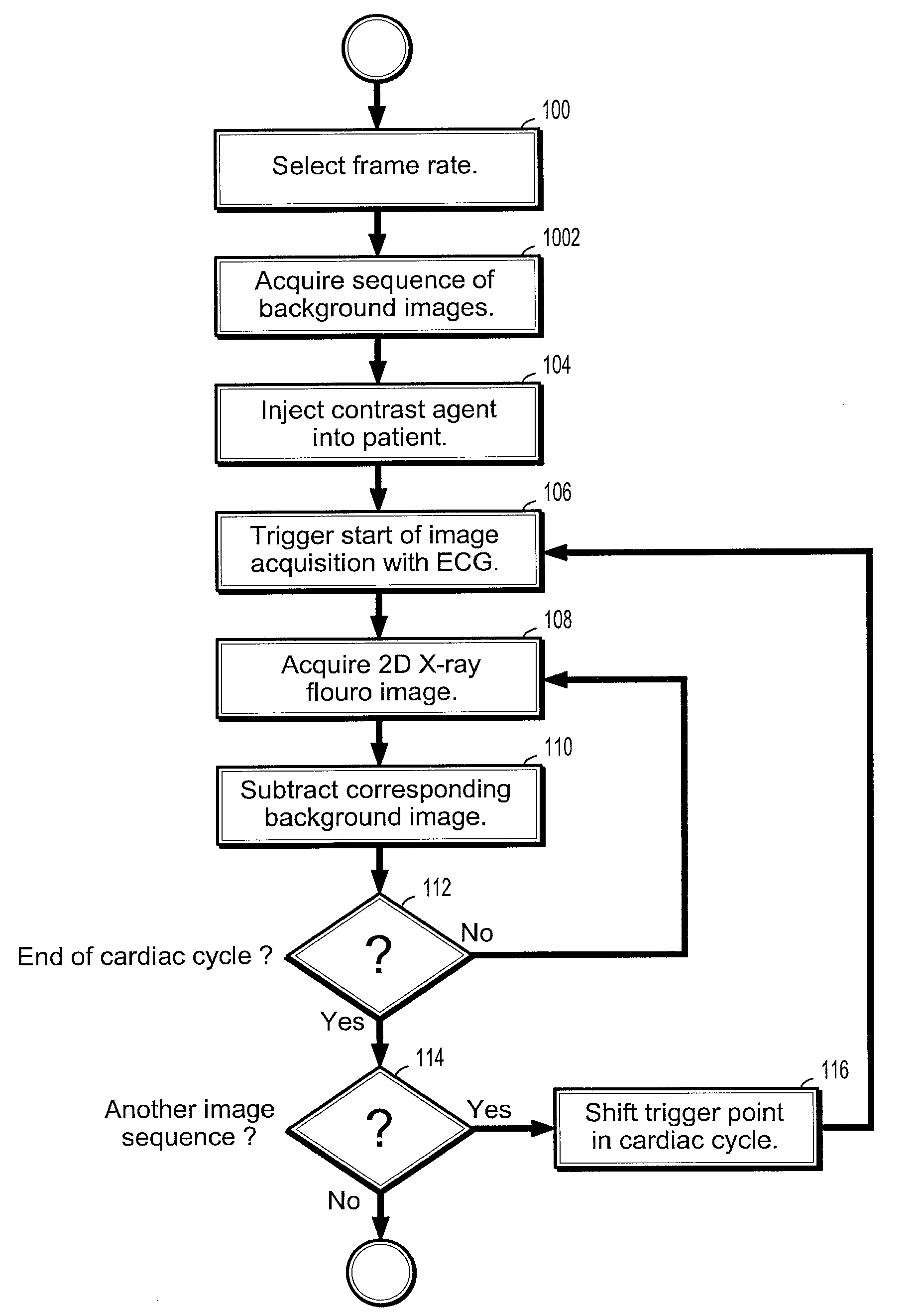
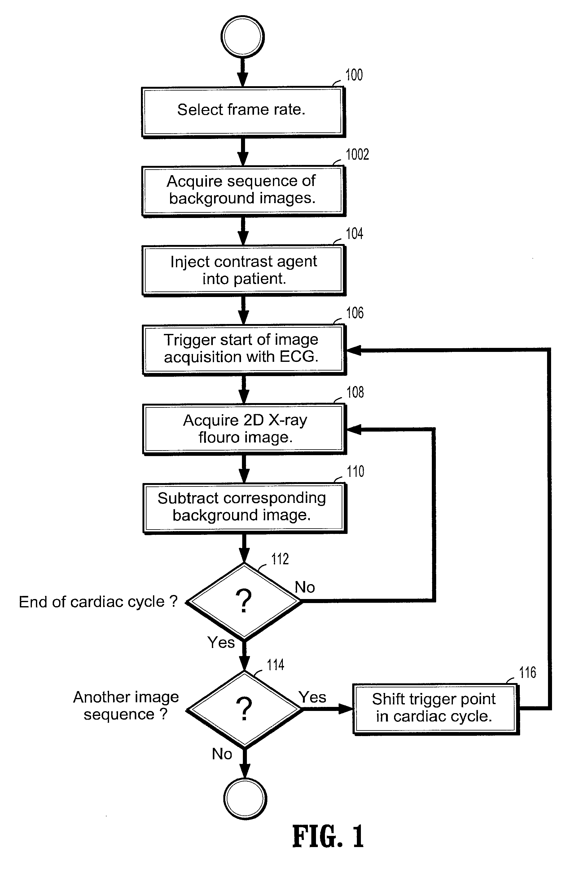
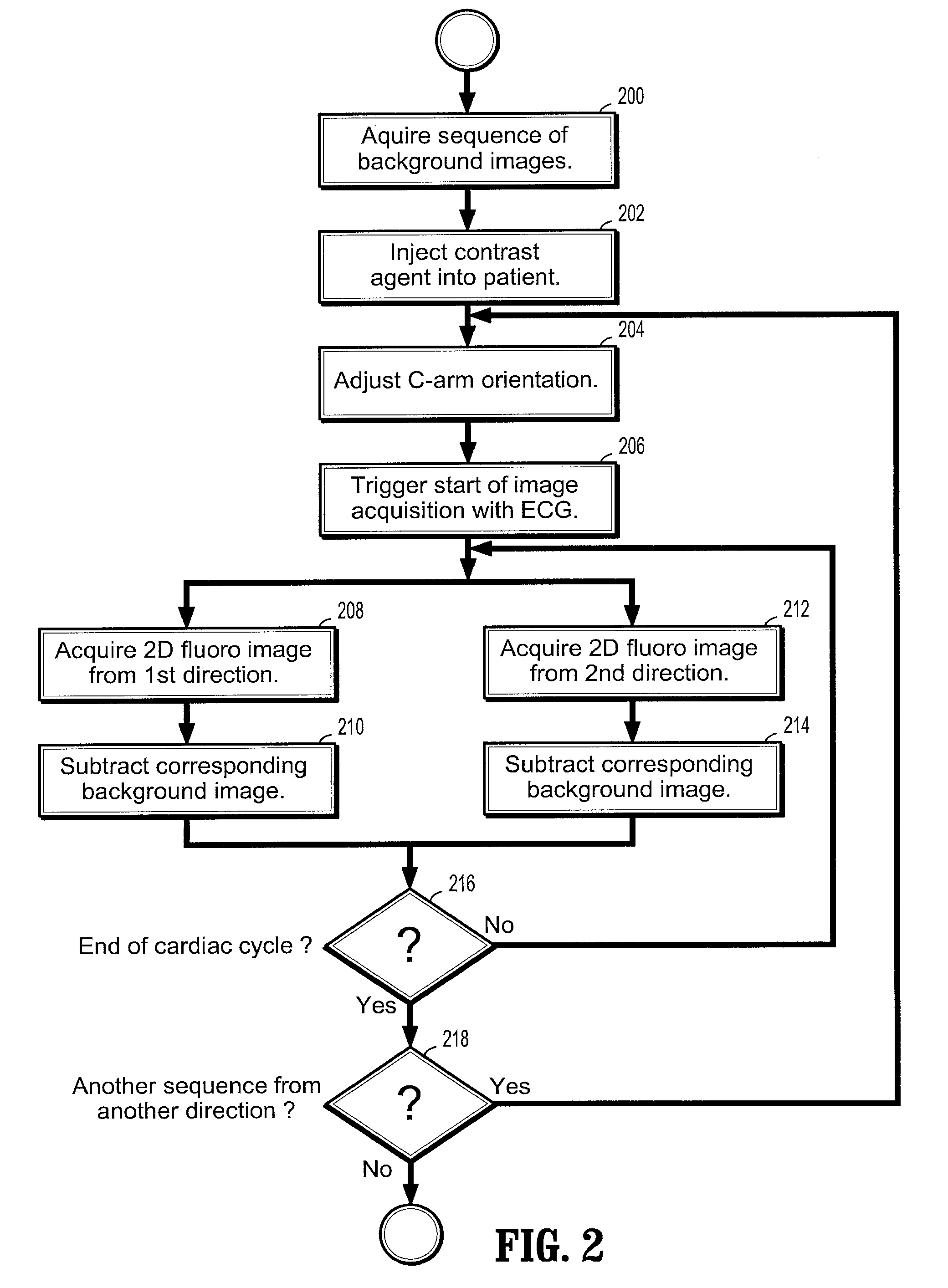
[0029]According to another aspect of the invention, there is provided a method of assessing rupture risk of an aneurysm from time-resolved images including injecting a contrast enhancing agent into a patient with an aneurysm, using an electrocardiogram (ECG) signal to trigger an image acquisition run, wherein a sequence of 2D X-ray fluoroscopic images is acquired, along with a corresponding ECG signal value, rotating a C-arm attachi

Problems solved by technology

Rupture and blood clotting are the risks involved with aneurysms.
This bulge in a blood vessel can burst and lead to death at any time, much like a bulge in an over-inflated innertube.
Rupture leads to drop in blood pressure, rapid heart rate, and lightheadedness.
The risk of death is high except for rupture in the extremities.
The larger an aneurysm becomes, the more likely it is to burst.
Pseudoaneurysms can be due to trauma involving the intima of the blood vessel, and are a known complication of percutaneous arterial procedures.
The lower extremities have higher arterial resistance, and the repeated trauma of a reflected arterial wave on the distal aorta may injure a weakened aortic wall and contribute to aneurysmal degeneration.
As tension increases, risk of rupture increases.
Increased pressure, such as that due to systemic hypertension, and increased aneurysm size aggravate wall tension and therefore increase the risk of rupture.
However, the correct modeling of the flow inside a vessel is impossible to validate in-vivo.
In addition, patient-specific modeling of the mechanical properties of the vessel walls cannot be done at all based on the 3D Angiography or CTA images.

Method used


Image

  • System and method for x-ray based assessment of aneurysm pulsation
  • System and method for x-ray based assessment of aneurysm pulsation
  • System and method for x-ray based assessment of aneurysm pulsation

Examples


Embodiment Construction

[0039]Exemplary embodiments of the invention as described herein generally include systems and methods for time-resolved imaging of pulsating aneurysms. Accordingly, while the invention is susceptible to various modifications and alternative forms, specific embodiments thereof are shown by way of example in the drawings and will herein be described in detail. It should be understood, however, that there is no intent to limit the invention to the particular forms disclosed, but on the contrary, the invention is to cover all modifications, equivalents, and alternatives falling within the spirit and scope of the invention.

[0040]As used herein, the term “image” refers to multi-dimensional data composed of discrete image elements (e.g., pixels for 2-D images and voxels for 3-D images). The image may be, for example, a medical image of a subject collected by computer tomography, magnetic resonance imaging, ultrasound, or any other medical imaging system known to one of skill in the art. T...


PUM


Abstract

A method of assessing rupture risk of an aneurysm from time-resolved images includes injecting a contrast enhancing agent into a patient with an aneurysm, using an electrocardiogram (ECG) signal to trigger an image acquisition run, wherein a sequence of 2D X-ray fluoroscopic images is acquired along with a corresponding ECG signal value, rotating a C-arm attaching the X-ray fluoroscopic imaging apparatus during said image acquisition run, wherein the images in said X-ray fluoroscopic image sequence are acquired from a rotating viewpoint, sorting the images in said X-ray fluoroscopic image sequence into time windows of the cardiac cycle based on the ECG signal, and constructing one or more 3-dimensional (3D) angiography image volumes of said aneurysm from said 2D fluoroscopic image sequence.

Description

CROSS REFERENCE TO RELATED UNITED STATES APPLICATIONS[0001]This application claims priority from: “X-ray based assessment of aneurysm pulsation”, U.S. Provisional Application No. 60 / 794,718 of Sauer, et al., filed Apr. 24, 2006, the contents of which are incorporated herein by reference.TECHNICAL FIELD[0002]This disclosure is directed to real-time X-ray fluoro imaging, in particular to time-resolved imaging of pulsating aneurysms.DISCUSSION OF THE RELATED ART[0003]An aneurysm (or aneurism) is localized, blood-filled dilation (bulge) of a blood vessel caused by disease or weakening of the vessel wall. Aneurysms can occur anywhere where there is a blood vessel, although they are most common in arteries. Aneurysms most commonly occur in arteries at the base of the brain, in the circle of Willis, and in the aorta, the main artery coming out of the heart. Rupture and blood clotting are the risks involved with aneurysms. This bulge in a blood vessel can burst and lead to death at any time...

Claims


Application Information

Patent Timeline
IPC IPC(8): A61B6/00G06K9/00
CPCA61B6/4441A61B6/503A61B6/504A61B6/541G06T2207/30101G06T7/0012G06T7/602G06T2207/10116G06T5/50G06T7/62
Inventor HALL, ANDREW F.MOHAMED, ASHRAFMOORE, TERIROHM, ESTHERSAUER, FRANK
Owner SIEMENS MEDICAL SOLUTIONS USA INC
Learn More