
Microsurgical injection and/or distending instruments and surgical method and apparatus utilizing same

a technology of microsurgical injection and distending instruments, which is applied in the field of microsurgical injection and/or distending instruments and surgical methods and apparatuses utilizing same, and can solve the problems of irreversible vision loss, severe vision loss, and neovascular glaucoma, and none of them have proved to be effectiv

Inactive Publication Date: 2003-09-11
PAQUES MICHEL +1
View PDF4 Cites 63 Cited by
  • Summary
  • Abstract
  • Description
  • Claims
  • Application Information

AI Technical Summary

Benefits of technology

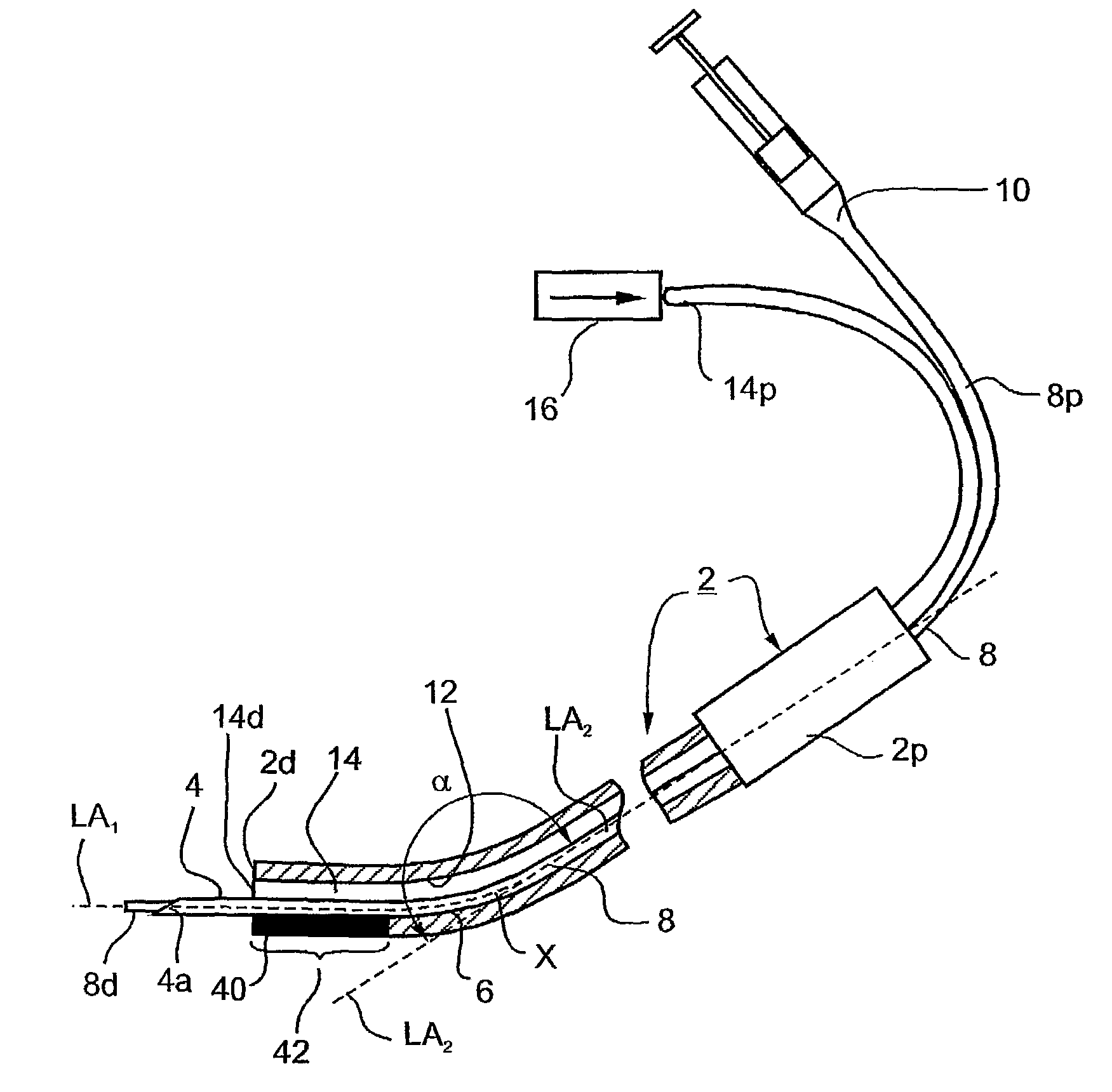
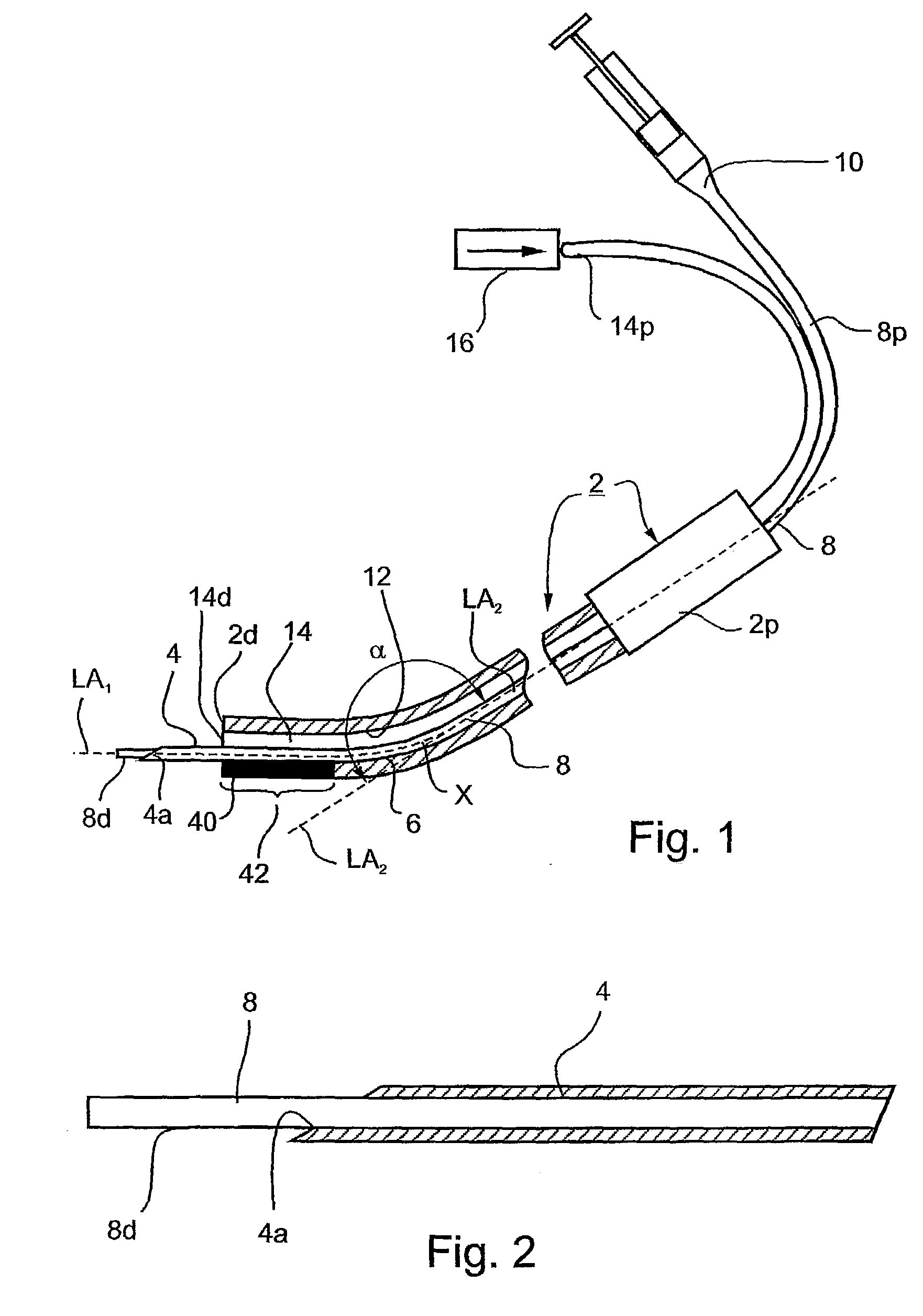
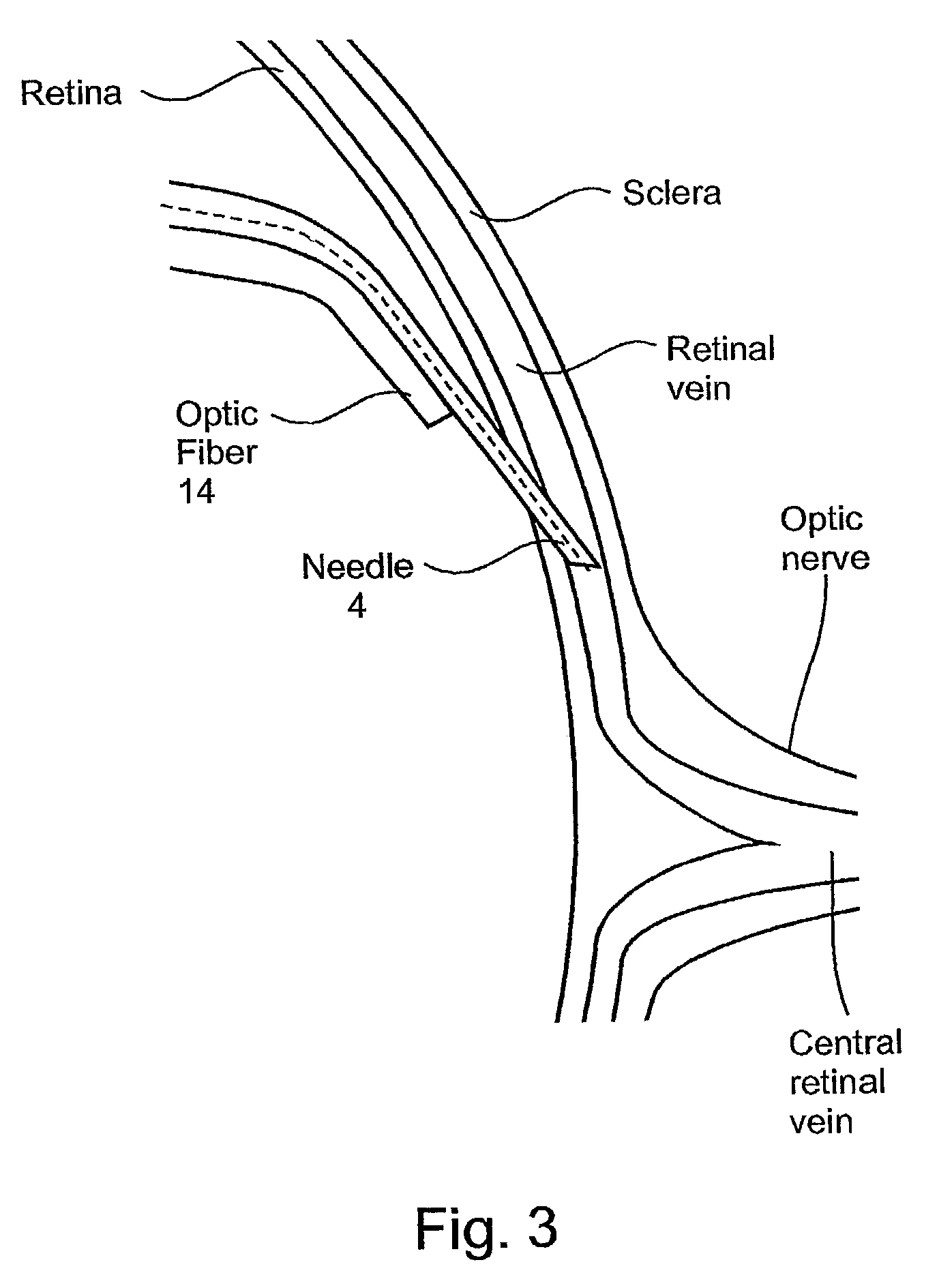
[0017] According to one aspect of the present invention, there is provided a microsurgical injection instrument particularly useful by a physician for injecting a substance into a blood vessel in the retina of a subject's eye, comprising: a hand piece having a proximal end graspable by the physician, and a distal end carrying a hollow miniaturized needle sharpened at its tip for penetrating the blood vessel in the subject's retina; the handpiece being formed with at least one passageway there through from its proximal end to the hollow needle at the distal end for the delivery thereto of the substance to be injected; the longitudinal axis of the distal end of the handpiece and of the hollow needle being an angle (".alpha.") of from 90.degree.-180.degree., preferably about 120.degree.-170.degree., most preferably about 145.degree., to the longitudinal axis of the proximal end of the handpiece, to facilitate orienting the needle coaxial with the retinal vein in the subject's retina and substantially tangentially to the plane of the subject's retina and thereby to facilitate penetrating the blood vessel in the subject's retina.
[0020] Another improvement of the device includes a plate that is placed under the needle to be apposed at the surface of the retina, which plate is useful for stabilizing the needle by formation of contact between the retinal surface and the device during the penetration of a vessel by the miniaturized needle. Then, the plate located under the miniaturized needle at the distal end is apposed against the retina to improve the stability of the device during the penetration of the vessel.
[0026] According to a further feature in the latter embodiment, the distal end of the handpiece of the microsurgical illumination instrument is constructed so as to be engageable with the distal end of the microsurgical injection instrument for stabilizing and guiding the distal end of the injection instrument when inserted into the subject's eye.

Problems solved by technology

Central retinal vein occlusion (CRVO) is an acute occlusion of the central retinal vein of the eye and can lead to a severe decrease of vision.
CRVO can also cause permanently damaging complications such as macular edema, one of the leading causes of visual loss in retinal pathology, and retinal ischemia, which can lead to irreversible loss of vision and neovascular glaucoma.
Many treatments such as troxerutin, heparin, hemodilution, laser photocoagulation have been proposed, but none has proved to be effective, and none is used in current practice.
However, it has been found that creating chorio-retinal anastomosis require high energy laser that can lead to unacceptable eyes complications such as choroidal and retinal neovascularization or vitreous hemorrhage.
However, several complications such as hemiplegia or even fatal stroke have been described in those studies.
Such risks inherent to injection of fibrinolytic in the general circulation are unacceptable for a non-life-threatening condition such as retinal vein occlusion.
However it remains a heavy procedure and the fibrinolytic agent is not delivered directly into the retinal vein.
Glass micropipettes are fragile and can easily be broken within the eye or within the retinal vein during the surgical procedure.
This risk makes the procedure unsafe.
Also, the external manipulator needed to stabilize the needle placed in the vessels in the XYZ axis makes the procedure cumbersome.
However, none of these known instruments appears to be suitable for the above treatment of venous occlusive diseases.

Method used


Image

  • Microsurgical injection and/or distending instruments and surgical method and apparatus utilizing same
  • Microsurgical injection and/or distending instruments and surgical method and apparatus utilizing same
  • Microsurgical injection and/or distending instruments and surgical method and apparatus utilizing same

Examples


Embodiment Construction

[0039] The microsurgical injection instrument illustrated in FIG. 1 is particularly useful by a physician for the treatment of retinal diseases, especially central retinal vein occlusion (CRVO) or branch retinal vein occlusion (BRVO), as briefly described above, by reestablishing retinal blood flow by pharmacological and mechanical means, by injecting a liquid substance or suspension, particularly a fibrinolytic agent into a blood vessel, and / or by catheterizing the blood vessel, in the retina of a subject's eye.

[0040] The illustrated instrument includes a handpiece, generally designated 2, of rigid material, plastic or metal. It has a finger-piece 2a at its proximal end 2p graspable by the physician, and a distal end 2d carrying a hollow needle 4 sharpened at its tip 4a for penetrating a blood vessel in the subject's retina. As will be described more particularly below, when the illustrated instrument is used for treating for CRVO or BRVO, the blood vessel penetrated would be a ret...


PUM


Abstract

A microsurgical injection instrument particularly useful by a physician for injecting a liquid substance or a suspension into a blood vessel in the retina of a subject's eye, includes a handpiece carrying a hollow needle at its distal end, for penetrating the blood vessel. The longitudinal axis of the distal end of the handpiece and of the needle is at an angle of from 90°-180°, preferably about 145°, to the longitudinal axis of the proximal end of the handpiece to facilitate orienting the needle substantially tangentially to the plane of the subject's retina coaxially to a blood vessel for penetrating the blood vessel. The instrument may also be used to treat an occluded blood vessel by moving a flexible tube through the hollow needle, after the needle has penetrated the blood vessel, to enter and catheterize the blood vessel.

Description

FIELD AND BACKGROUND OF THE INVENTION[0001] The present invention relates to microsurgical instruments and also to a surgical method and apparatus utilizing such instruments. One embodiment of the invention is particularly useful for injecting a liquid or a suspension substance into a blood vessel in the retina of a subject's eye in order to treat certain eye diseases, such as retina disease, therein, and is therefore described below with respect to this application. Another embodiment of the invention is useful for catheterizing (or distending or cannulating) an occluded blood vessel, such as in a subject's eye, and is therefore described below also with respect to this application.[0002] Venous occlusive diseases are among the most common retinal diseases seen in clinical practice. Recognition of these diseases is of particular importance because their complication may cause significant visual morbidity.[0003] Central retinal vein occlusion (CRVO) is an acute occlusion of the cent...

Claims


Application Information

Patent Timeline
IPC IPC(8): A61B19/00A61F9/00
CPCA61B2019/5206A61F2009/0052A61F9/0017A61B2090/306
Inventor PAQUES, MICHELBENHAMOU, NATHANAEL
Owner PAQUES MICHEL
Learn More