Automatic contouring of tissues in CT images

a tissue contouring and tissue technology, applied in image enhancement, image analysis, instruments, etc., can solve the problems of insufficient robustness of clinical use, inexact method, prostate cancer,

Inactive Publication Date: 2004-05-27
NORTHROP GRUMAN CORP
View PDF3 Cites 42 Cited by
  • Summary
  • Abstract
  • Description
  • Claims
  • Application Information

AI Technical Summary

Problems solved by technology

This outlining procedure is referred to as contouring and is a very lengthy, inexact method, especially when employed with certain types of cancer.
For example, a particularly prominent form of cancer in males, prostate cancer, is problematic in this regard because of the close proximity of the prostate to other organs including the bladder, the rectum and the seminal vesicles, and because of the nearly uniform density of these tissues to X-rays.
Unfortunately, the prior art approaches to such autocontouring techniques employ 2D and 3D region growing, which is an area-based technique.
This approach has not been found robust enough for clinical use and has no current commercial application.
This can happen, for example, when the object being contoured is not uniform in appearance.

Method used


Image

  • Automatic contouring of tissues in CT images
  • Automatic contouring of tissues in CT images
  • Automatic contouring of tissues in CT images

Examples


Embodiment Construction

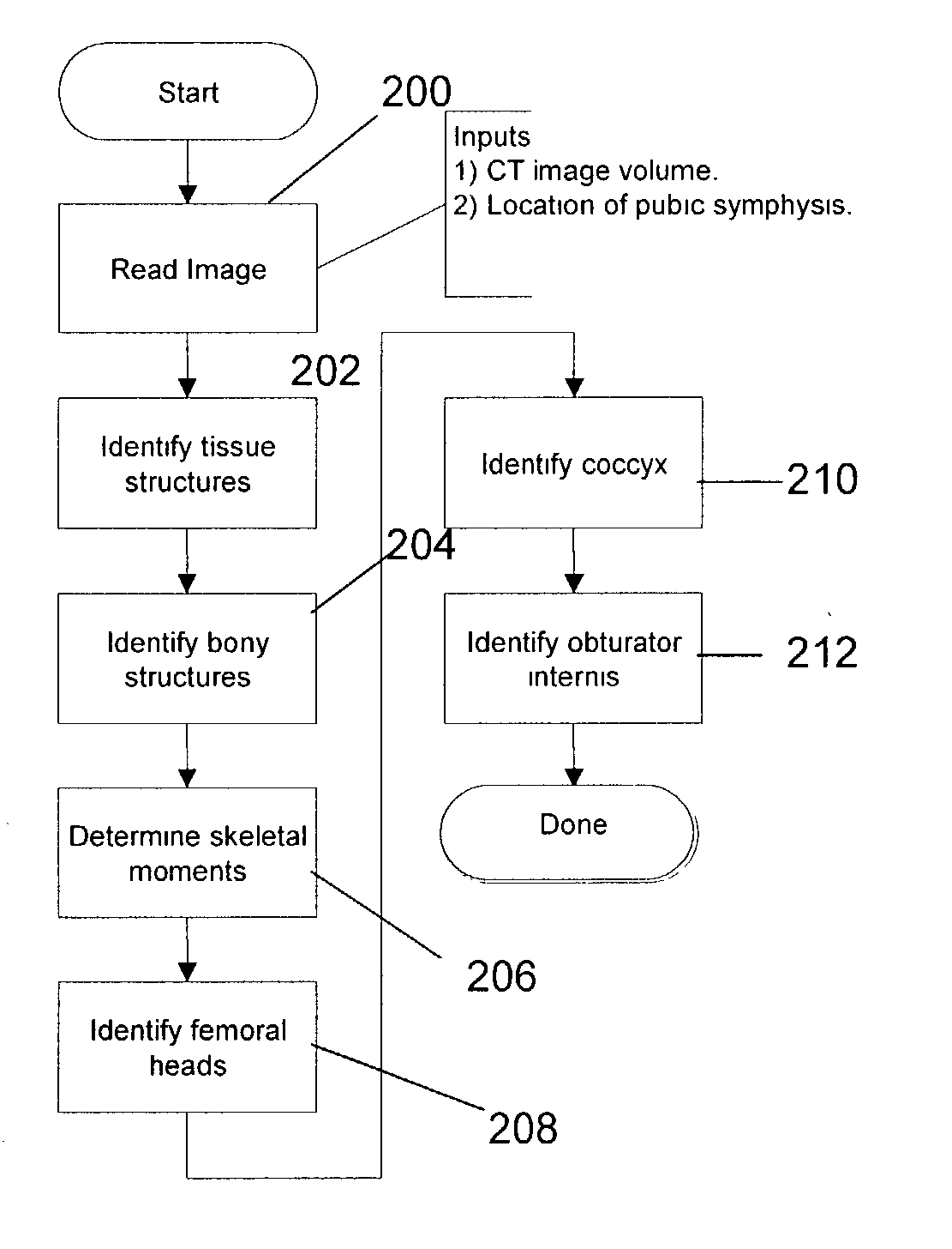
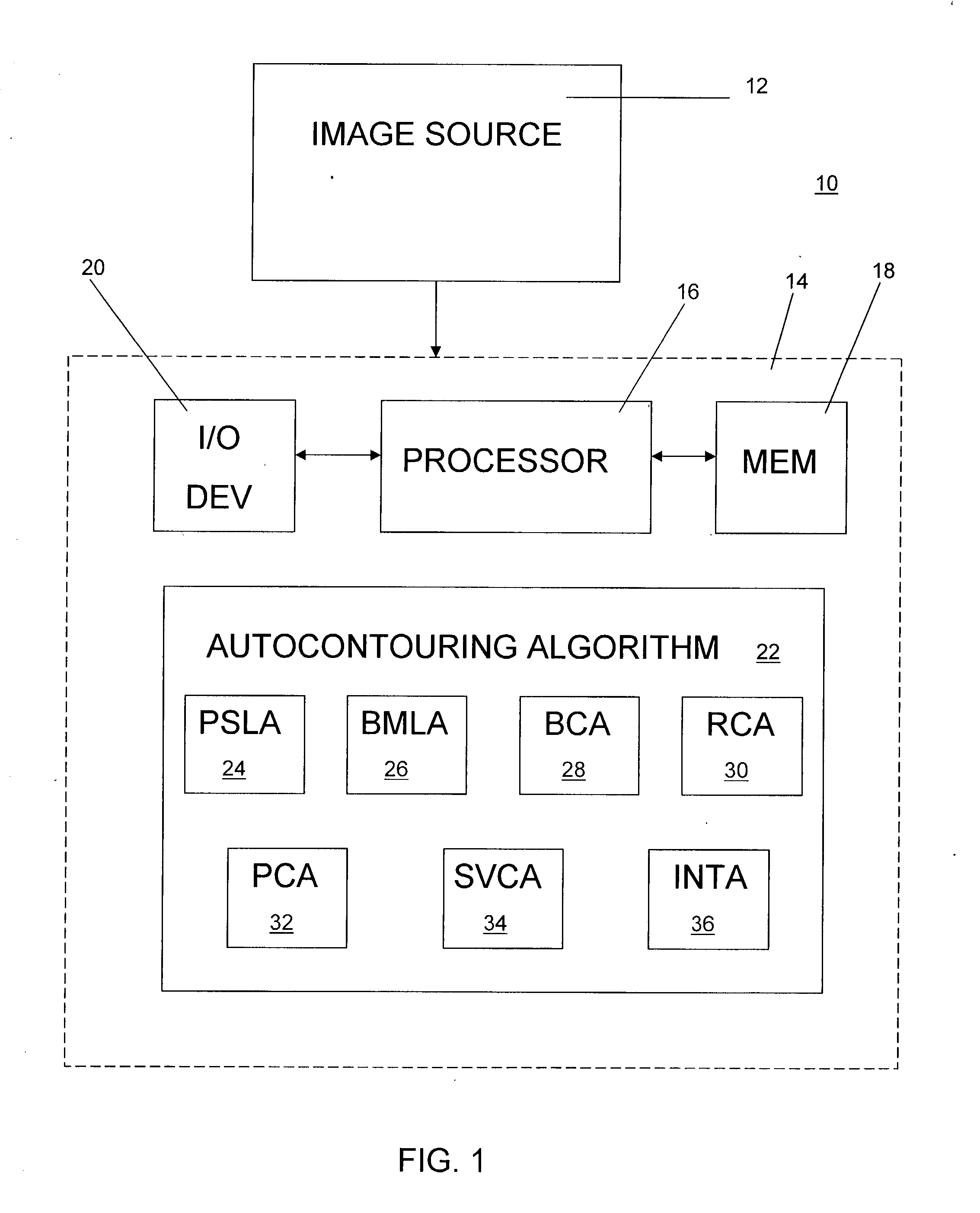
[0022] FIG. 1 illustrates a CT image analysis system 10 for generating and contouring 3D volume images in accordance with a preferred embodiment of the present invention. The preferred embodiment is specifically designed to contour the organs in a male's pelvic region, although it should be understood that the invention could also be employed to contour images of other anatomic organs or structures. In addition, although the preferred embodiment is designed specifically for analyzing CT images, it should be understood that the invention could also be employed for analyzing other types of medical images, such as MRI, ultrasound, etc.

[0023] In the preferred embodiment, the system 10 includes a source 12 of CT 3 -dimensional volume images of a person's body. The source 12 can be any suitable system or device for storing CT images, such as for example, a remote network or database that may be accessed in any known manner, such as over the Internet, or a removable storage device. As is c...


PUM


Abstract

An automated method and system for autocontouring organs and other anatomical structures in CT and other medical images employs one or more contouring techniques, depending on the particular organs or structures to be contoured. In a preferred embodiment, an edge-based technique is employed to contour one or more organs. A multiple hypothesis testing technique can be employed to improve the accuracy of the resulting contour. Independent algorithms can be employed for contouring multiple organ in a given region, such as the male pelvic region. An integration algorithm can be employed to combine the results of the independent algorithms to improve accuracy further.

Description

[0001] 1. Field of the Invention[0002] The present invention relates in general to a method and system for automatically contouring tissues and other anatomic structures in CT or other medical images. The method and system employ algorithms using edge detection and other techniques to identify boundaries of anatomic structures, such as organs.[0003] 2. Description of the Background Art[0004] Radiation treatment is often used to treat various forms of cancer. Modern radiation treatment techniques, such as intensity-modulation radiation therapy (IMRT), are capable of preferentially concentrating radiation in specific cancerous tissues in the body while limiting damage to nearby normal tissues. However, in order for this these techniques to be effective, a physician or other highly trained individual must accurately identify which tissues are to be treated and which are to be avoided. As a result, current methods of radiation treatment planning require the physician or other highly tra...

Claims


Application Information

Patent Timeline
Patent Type & Authority Applications(United States)
IPC IPC(8): G06K9/00G06T5/00G06T7/00G06T7/60
CPCG06T7/0012G06T7/0083G06T2207/30008G06T2207/10081G06T2207/20168G06T7/606G06T7/12G06T7/66
Inventor SIVARAMAKRISHNA, RADHIKABIRBECK, JOHN S.FRIELER, CLIFF E.COTHREN, ROBERT M.
Owner NORTHROP GRUMAN CORP
Why Eureka
Try Eureka