
Image processing apparatus and image processing method

a technology of image processing and apparatus, applied in the field of image processing apparatus and a method, can solve the problems of difficult to extract the boundary of the cardiac muscle automatically, difficult to detect a low luminance change boundary, and difficult to extract the endocardium and the epicardium accurately, stably and automatically

Inactive Publication Date: 2004-12-09
KK TOSHIBA
View PDF53 Cites 17 Cited by
  • Summary
  • Abstract
  • Description
  • Claims
  • Application Information

AI Technical Summary

Problems solved by technology

Artifacts, noise, etc. superposed on the images, however, make it difficult to extract the boundaries of the cardiac muscle automatically.
When change in luminance is used, it is however difficult to detect a boundary low in luminance change.
For this reason, it is difficult to extract both the endocardium and the epicardium accurately, stably and automatically.
Even in this technique, sufficient accuracy cannot be obtained because there is the possibility that the foot of the luminance distribution may be extended while it exceeds n % of the maximum luminance when luminance change in a boundary and its vicinity is obscure.
If a predetermined range is entirely used for calculation based on the temporary boundaries in order to search for the combination, the volume of calculation becomes huge.
It was however difficult to detect the outer boundary of the heart wall stably because luminance change in the outer boundary of the heart wall was obscure.
As a result, detection of the thickness of the heart wall was apt to be unstable because of the influence of fluctuation caused by obscurity.

Method used

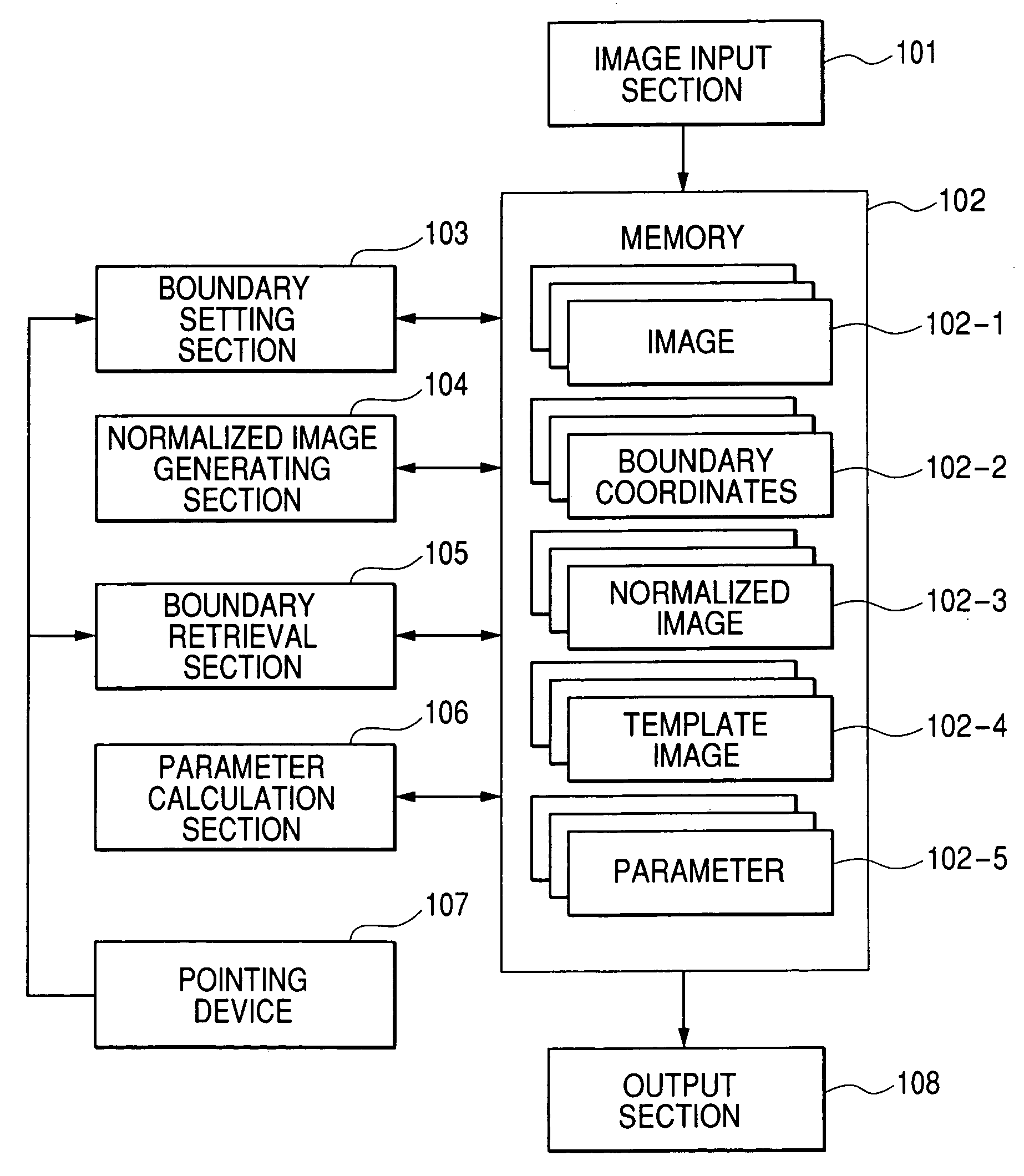
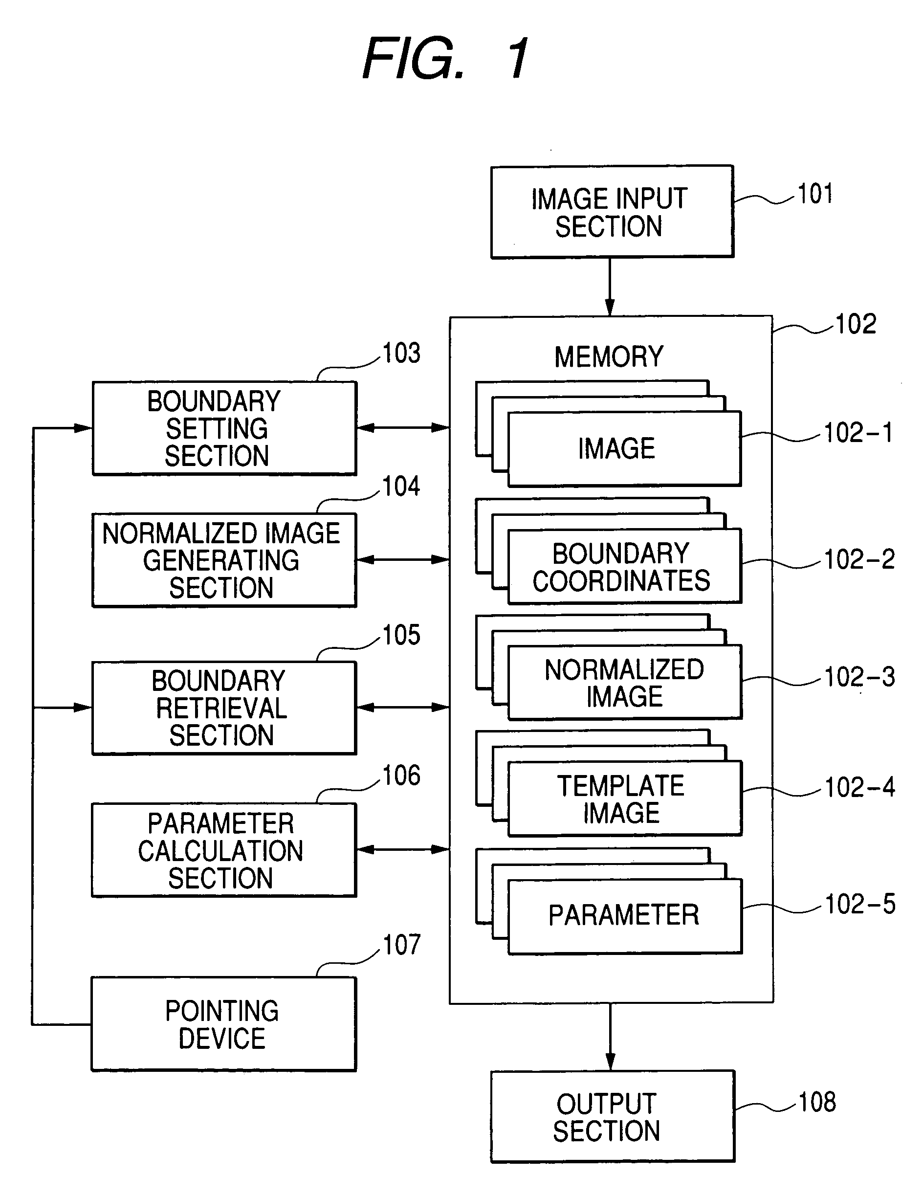
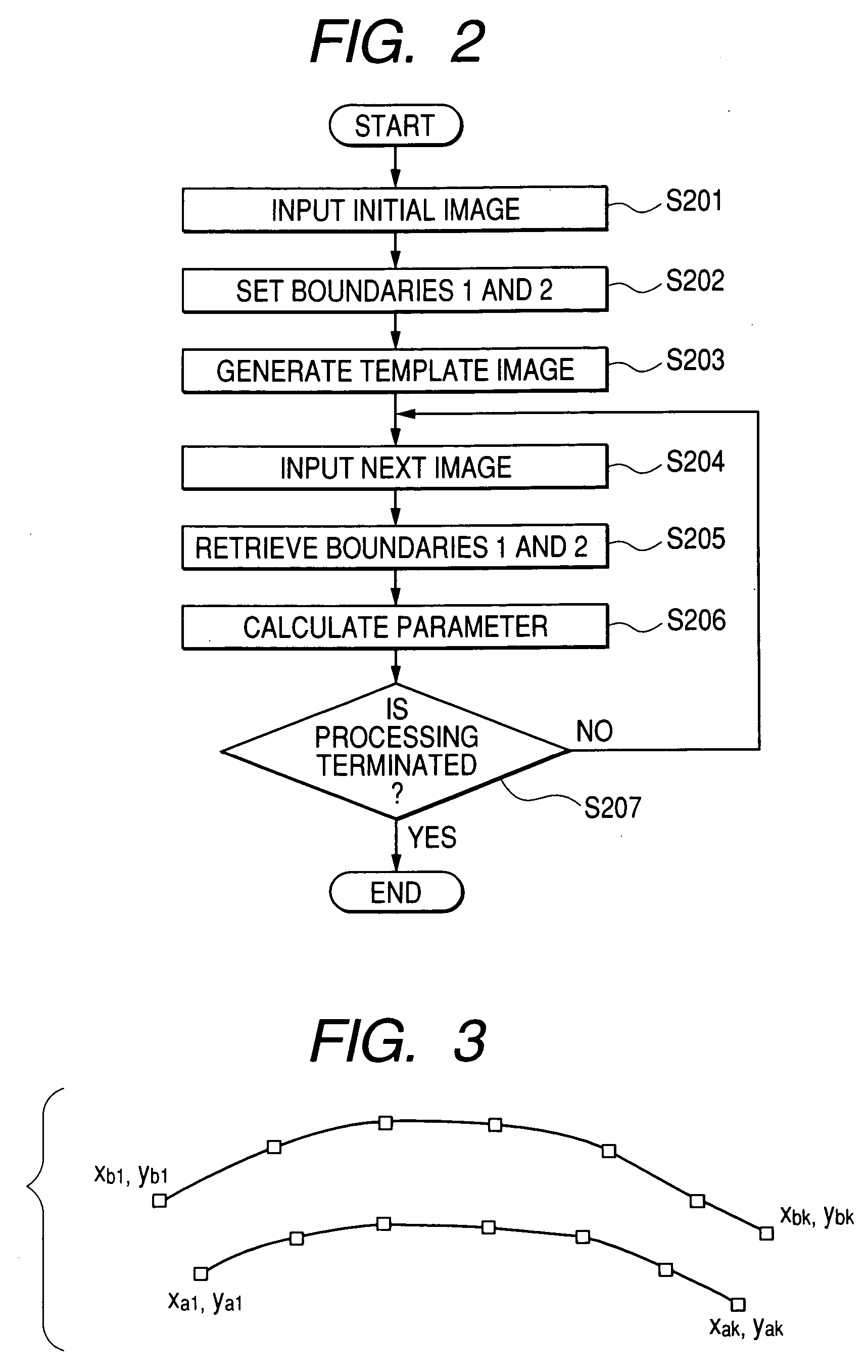

Image

  • Image processing apparatus and image processing method
  • Image processing apparatus and image processing method
  • Image processing apparatus and image processing method

Examples


Embodiment Construction

of Retrieval Method has shown the case where a subject of measurement of wall thickness change of the heart wall is divided into segments so that points expressing the boundary 2 are set for the boundaries of each segment, the boundaries may be expressed by smaller number of parameters according to an interpolating method using a splined curve or the like.

[0116] Although the description has been made while the heart is taken as an example, thickness change of a wall or the like can be also detected in other internal organs (such as the stomach, the liver, the urinary bladder, the kidney and the uterus) and an unborn baby as well as the heart.

[0117] (Modification) This embodiment may be provided as a mode for a medical work station containing not only a process of measuring wall thickness change of the heart wall but also other image measuring process and a data management function such as filing.

[0118] Alternatively, a series of heart wall thickness change measuring steps for image ...


PUM


Abstract

A boundary 1 of a wall in an initial image of time-series images is decided automatically or manually. A boundary 2 is generated automatically or manually in a neighbor of the other boundary of the wall. A normalized image is generated on the basis of an image pattern of a region surrounded by the boundaries 1 and 2 and registered as a template image. In each of the time-series images, boundaries 1 and 2 that generate a normalized image most similar to the template image are calculated. In this manner, wall thickness change can be calculated automatically stably and accurately even in the case where luminance change in the boundaries of a wall is obscure. This method is useful for diagnosis of heart disease etc. in medical image diagnosis.

Description

[0001] This application is based upon and claims the benefit of priority from the prior Japanese Patent Application No.2003-60876 filed on Mar. 7, 2003; the entire contents of which are incorporated herein by reference.[0002] 1. Field of the Invention[0003] The present invention relates to an image processing apparatus and a method for aiding diagnosis using medical images.[0004] 2. Description of the Related Art[0005] Change in thickness of cardiac muscle of a heart is a significant indicator in diagnosis of heart disease such as myocardial infarction.[0006] As a method for measuring change in thickness of cardiac muscle, there is a method including acquiring time-series cross-sectional images of a heart and its vicinity; detecting an inner boundary and an outer boundary of the cardiac muscle by image processing; and the calculating thickness of the cardiac muscle in accordance with each image.[0007] Artifacts, noise, etc. superposed on the images, however, make it difficult to ext...

Claims


Application Information

Patent Timeline
Patent Type & Authority Applications(United States)
IPC IPC(8): G01R33/54A61B5/00A61B5/055A61B6/00A61B8/00G06K9/00G06T1/00G06T5/00G06T7/00G06T7/60
CPCA61B6/503A61B8/0883G06T7/0012G06T7/0083G06T7/602G06T2207/10132G06T2207/20104G06T2207/30048Y10S128/922G06T7/12G06T7/62
Inventor NISHIURA, MASAHIDE
Owner KK TOSHIBA
Learn More