Three-dimensional tissue hardness imaging

a three-dimensional tissue and hardness imaging technology, applied in the field of three-dimensional tissue hardness imaging, can solve the problems of life-threatening thrombosis, difficult reproducible analysis of blood vessels in such a manner, and inability to obtain information from conventional echograms, etc., to achieve the effect of enabling detection of hardness and/or elasticity properties, optimizing correlation between consecutive images, and negative correlation

Inactive Publication Date: 2005-02-10
STICHTING VOOR DE TECH WETENSCHAPPEN
View PDF6 Cites 59 Cited by
  • Summary
  • Abstract
  • Description
  • Claims
  • Application Information

AI Technical Summary

Benefits of technology

[0009] The invention meets such needs and provides a method with which 3D information about the elasticity and / or hardness of a wall of a body cavity, in particular a blood vessel, can be obtained in a consistent and reproducible manner. In this regard, it is observed that with the present conventional technique the correlation between consecutive images is optimized by positioning the sensor as stably as possible, because movement of the sensor in general has a negative effect on the correlation. The invention is based on the insight that precisely by performing a motion transverse to the measuring plane a sufficient correlation between consecutive images can be maintained to enable detection of hardness and / or elasticity properties.
[0017] In the alternative or in addition thereto, at an assumed cyclic pressure change, signals can be received at predetermined time intervals in the period of the motion. In a preferred embodiment, these are signals of a blood vessel wall, the data being received only during a specific time interval of the period of the heartbeat. An advantage thereof is that signals that are not or less suitable for the determination of elasticity and / or hardness information of the tissue need not be stored, as a result of which data storage capacity can be performed to a limited extent, and the data processing can be significantly simplified.

Problems solved by technology

Such information is often not derivable from a conventional echogram, since the image of high-risk cannot be distinguished from less high-risk plaques.
If the stresses become too high, a plaque can tear open, so that a life-threatening thrombosis can arise.
With the present technique, it is practically very difficult to reproducibly analyze a blood vessel in such a manner.
Furthermore, on the basis of conventional echographic data it is very hard to localize a suspect spot in a blood vessel.
In fact, the performance of a single transverse scan at selected positions in a blood vessel provides insufficient information to enable determination of the presence or absence of plaques in the blood vessel as a whole.

Method used


Image

  • Three-dimensional tissue hardness imaging
  • Three-dimensional tissue hardness imaging
  • Three-dimensional tissue hardness imaging

Examples


Embodiment Construction

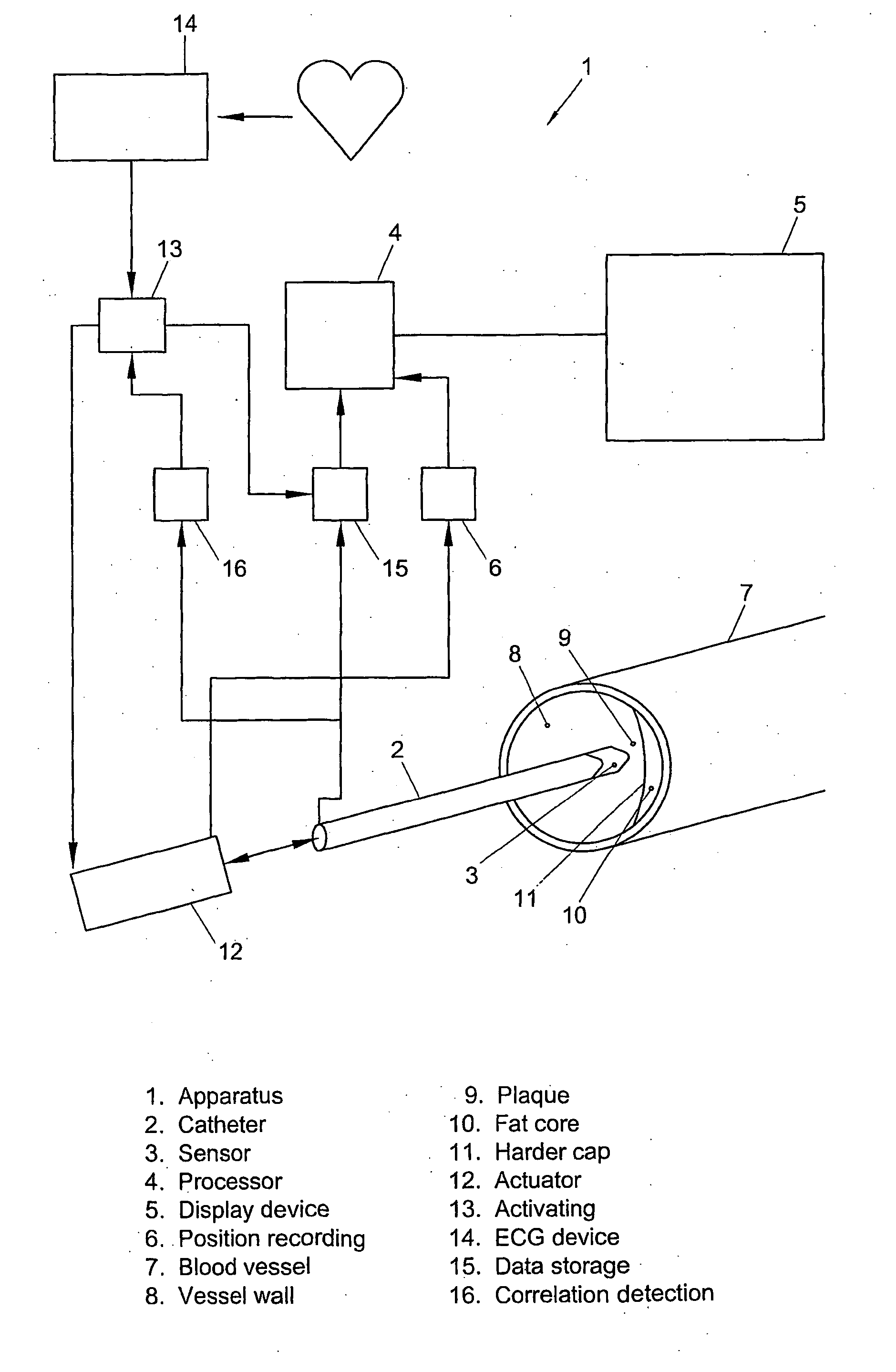
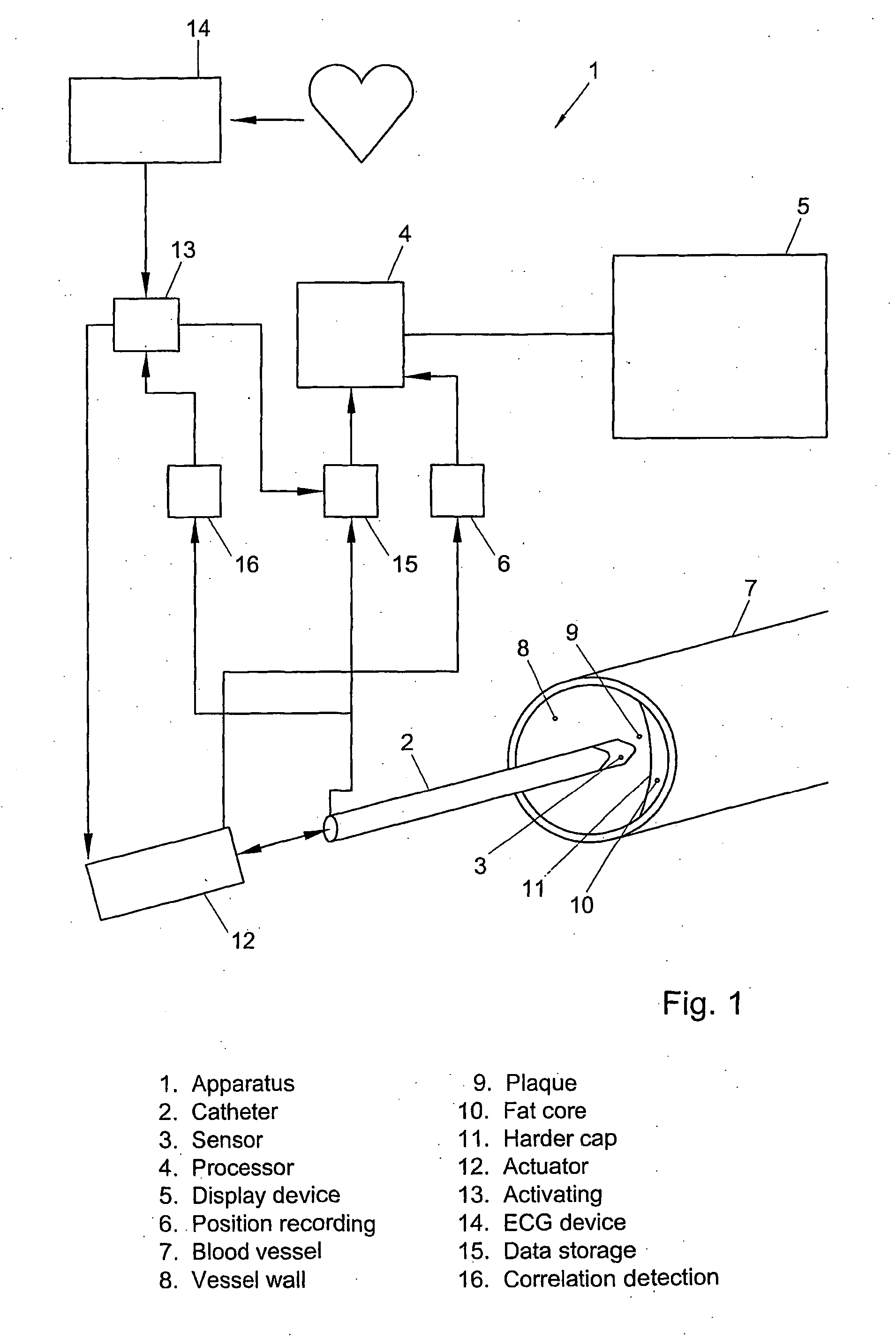
[0033]FIG. 1 is a diagrammatic representation of the apparatus 1 according to the invention. This apparatus comprises a movable catheter 2 provided with an acoustic sensor 3. A processor 4 is present to collect and process echographic data; the processor 4 is connected with a display device 5. The processor 4 is further in contact with a position recording means 6 for recording the position of the sensor 3.

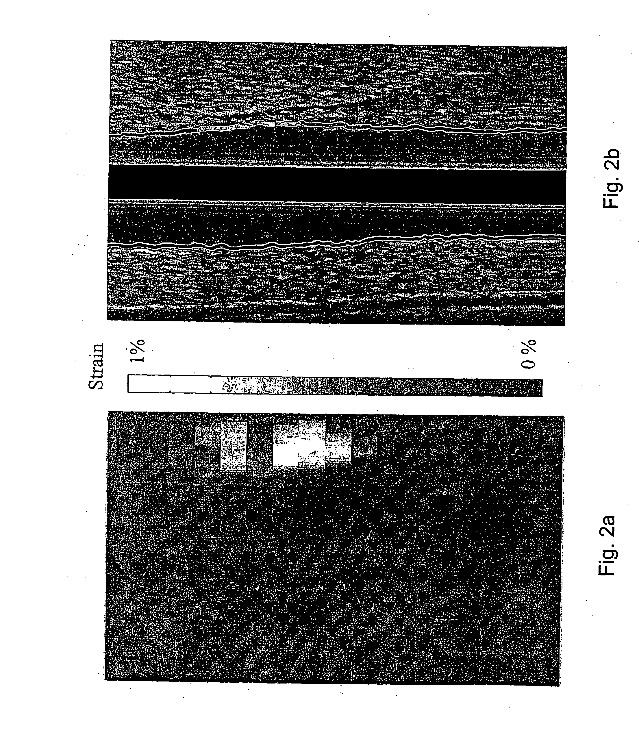
[0034] The catheter 2 can be moved through a blood vessel 7, which blood vessel 7 has a vessel wall 8 deformed by the heartbeat. The deformation can be derived by the processor 4 from the echographic data of the catheter 2 and related to elasticity and / or hardness parameters of the wall 8.

[0035] In explanation, a plaque 9 is shown in the blood vessel 7. This plaque comprises a fat core 10 closed by a harder cap 11. The motion of the catheter 2 is controlled by an actuator 12. The actuator 12 has an adjustable speed of motion, such that the catheter can be moved at a speed of 0.1...


PUM


Abstract

A method for generating hardness information of tissue subject to a varying pressure. The method comprises receiving signals from the tissue from a sensor for measuring the deformation of the tissue in a measuring plane defined by the sensor, which sensor, during a varying pressure exerted on the tissue, is moved along the tissue in a direction transverse to the measuring plane; identifying strain of the tissue from the resulting signals; and relating the strain to elasticity and / or hardness parameters of the tissue. The method may comprise the step of displaying elasticity and / or hardness parameters of a tissue surface or tissue volume part extending practically parallel to the direction of motion of the sensor.

Description

CROSS-REFERENCE TO RELATED APPLICATION [0001] This application is a continuation of PCT International Patent Application No. PCT / NL / 02 / 00572, filed on Sep. 2, 2002, designating the United States of America, and published, in English, as PCT International Publication No. WO 03 / 017845 A1 on Mar. 6, 2003, the contents of the entirety of which is incorporated by this reference.TECHNICAL FIELD [0002] This invention relates to a method for generating hardness information of tissue subject to a varying pressure. In particular, the method relates to a method for generating hardness information of the wall of a blood vessel or body cavity. BACKGROUND [0003] Such a method is known from European patent application EP-A 0 908 137. In this application, the strain (deformation) of vessel walls is derived with ultrasound from the relative displacement of a more inward layer and a more outward layer of the vessel wall as a result of the pressure varying through the heartbeat. These relative displac...

Claims


Application Information

Patent Timeline
Patent Type & Authority Applications(United States)
IPC IPC(8): A61B8/08A61B8/12
CPCA61B8/12A61B8/0891A61B8/485
Inventor VAN DER STEEN, ANTONIUS FRANCISCUS WILHELMUSDE KORTE, CHRISTOFFEL LEENDERTMASTIK, FRITSSCHAAR, JOHANNES ANTONIUS
Owner STICHTING VOOR DE TECH WETENSCHAPPEN
Why Eureka
Try Eureka