Tissue anchoring system and method

a tissue anchoring and tissue technology, applied in the field of surgical devices and methods, can solve the problems of increasing patient pain, long recovery time, difficult and impractical to make large access points, etc., and achieve the effect of maintaining the tension applied

Inactive Publication Date: 2005-02-10
ORTHODYNE
View PDF35 Cites 124 Cited by
  • Summary
  • Abstract
  • Description
  • Claims
  • Application Information

AI Technical Summary

Benefits of technology

[0012] The present invention also contemplates a bone opening protector having a first portion defining an opening therein to receive a cable. The first portion is adapted to engage a surface surrounding a bone opening. The protector also includes an anchoring extension, connected to the first portion, adapted to extend into bone to inhibit movement of the first portion.
[0013] The invention further contemplates a method for joining two sections of tissue. The method includes providing a first anchor, an elongated member attached to the first anchor, a second anchor slidable on the elongated member and having a deformable portion adapted to engage the elongated member, and a crimping tool having an outer member and an inner member movably disposed within the outer member. The first anchor is inserted into a first section of tissue. The second anchor is threaded on the elongated member. Tension is applied on the elongated member to urge the first section of tissue toward a second section of tissue. The elongated member is threaded through the crimping device, and the inner member of the crimping device is moved with respect to the outer member thereby deforming the deformable portion of the second anchor and maintaining the tension applied to the elongated member.

Problems solved by technology

The enlarged access site may increase patient pain and lengthen recovery time.
Further, in some locations it is difficult and impractical to make large access points to reach the appropriate site because of surrounding joints and vessels.
Further, some of these devices may be difficult to use since it may be hard to reduce a fracture between two bone segments and maintain that reduction while the device is inserted.
This is particularly true with small bone fragments were the use of threaded implants may tend to rotate one bone segment with respect to another, thereby creating a misalignment between the fragments.
A significant drawback of these systems is that they require access around the entire bone.

Method used


Image

  • Tissue anchoring system and method
  • Tissue anchoring system and method
  • Tissue anchoring system and method

Examples


first embodiment

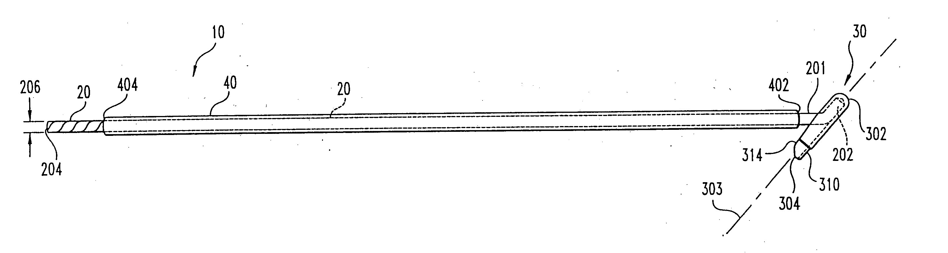
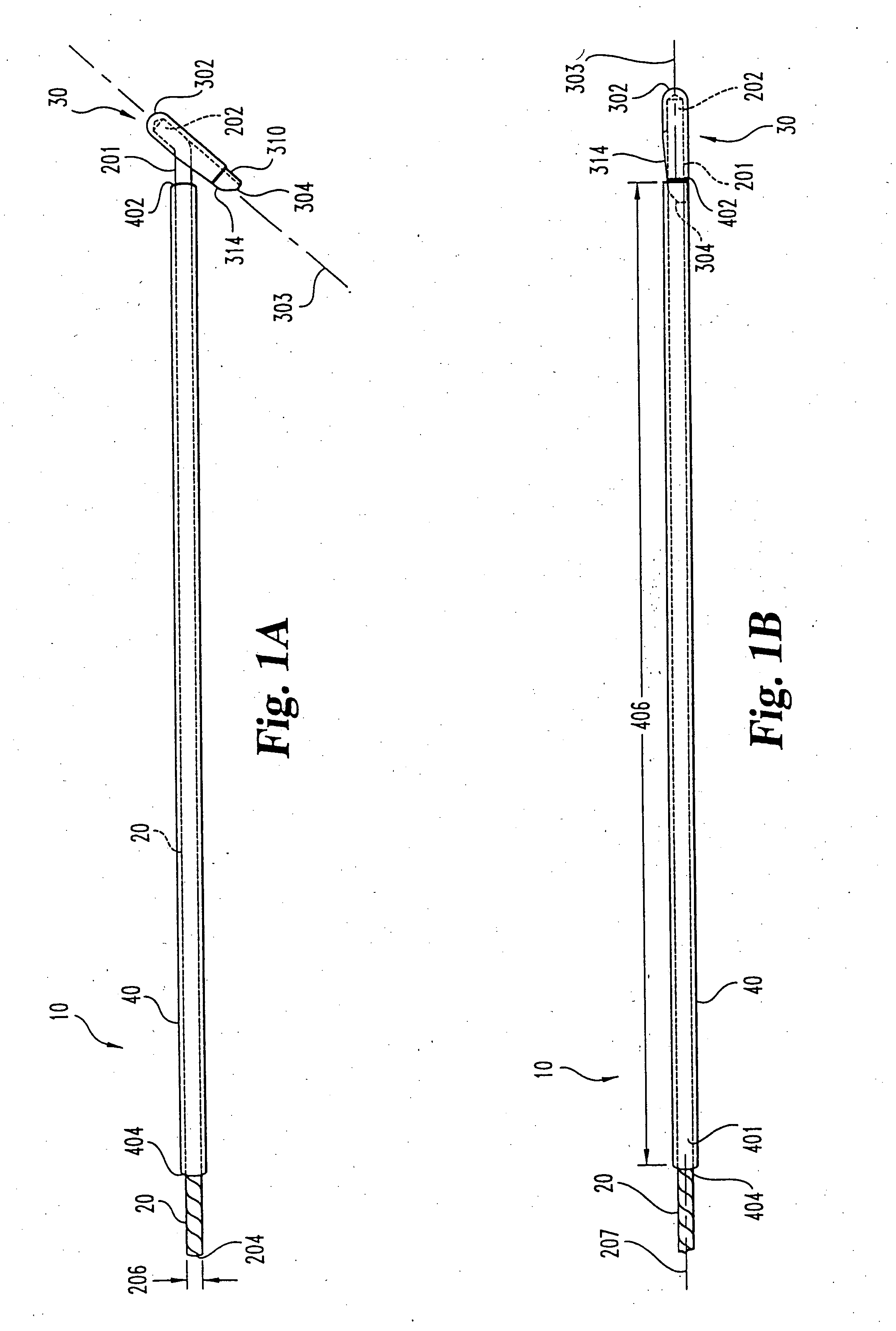
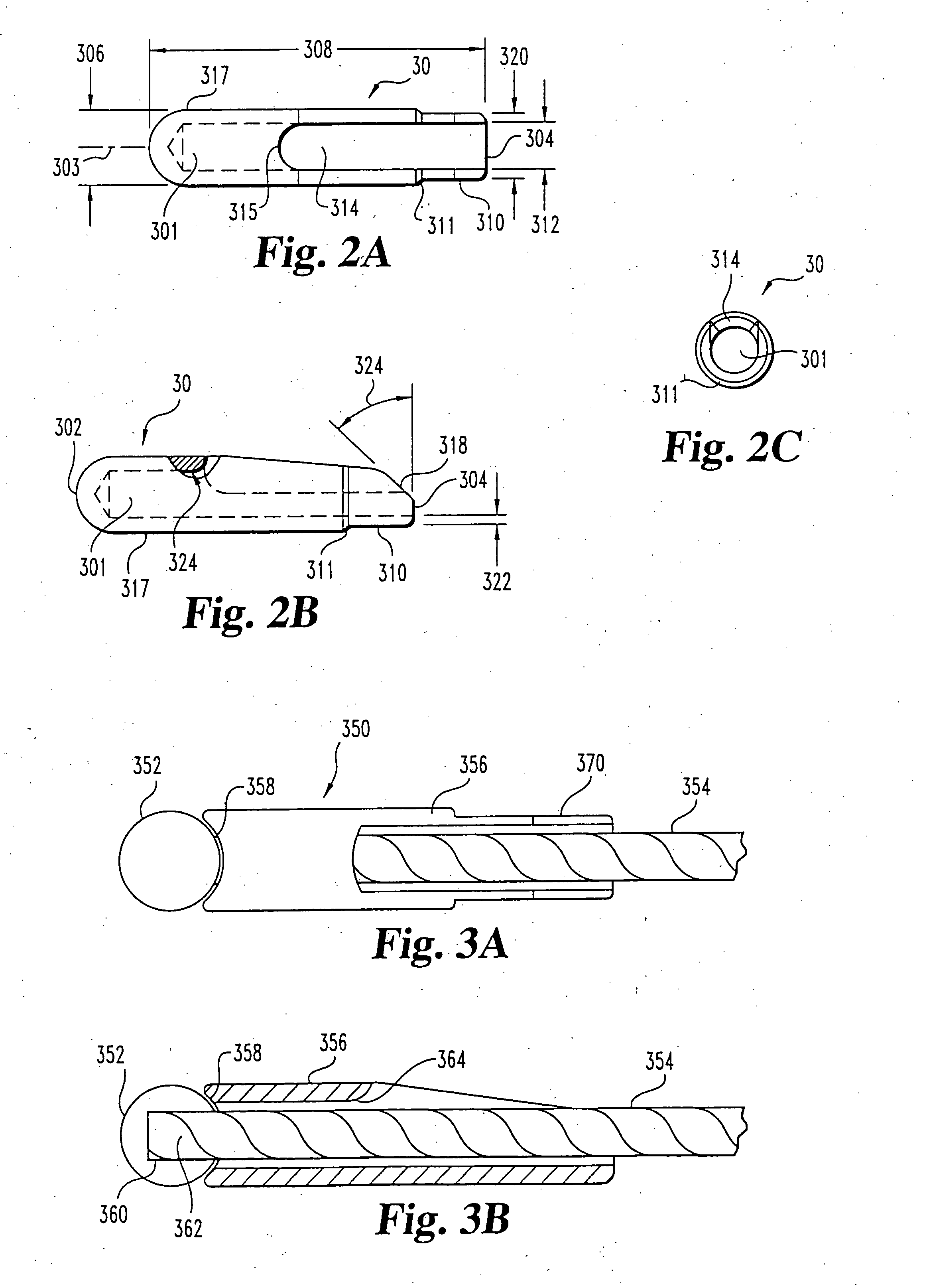
[0064] The present invention further contemplates a delivery device for insertion of a cable and anchor combination. Although the device is illustrated for use with the above described anchor, it is contemplated that the delivery device may be used or adapted for use with a variety of cable and anchor combinations. the delivery device 40 (FIGS. 1 and 2) comprises a rigid metallic cylindrical member having a length 405 sufficient to penetrate a desired surgical site and a longitudinal bore 401 extending from a proximal end 404 to a distal end 402. The bore 401 is sufficiently large to permit the anchor's proximal portion 310 to fit thereinto but too small to permit the anchor's distal portion 317 to fit thereinto. Although a preferred embodiment of the delivery device has the distal portion of anchor 30 extending distally beyond the distal end of the delivery device, it is contemplated that anchor 30 could be entirely retained within the delivery device until it is deployed. In this ...

second embodiment

[0066] In a delivery device according to the present invention, (FIGS. 4, 5A, and 5B), the impelling means comprises means for releasably maintaining a longitudinal force on cable 20 in a proximal direction, for retaining the anchor 30 in the first position.

[0067] In this embodiment, the delivery device 70 comprises a body 72, which has a handle portion 720 having a proximal end 722 and a recess 728 at the distal end 724. The recess 728, which extends from the top 721 to the bottom 723 of the delivery device body 72, is rounded at its proximal end 725. A longitudinal bore 726 extends from the proximal end 722 to the distal 724 end, the bore 726 being dimensioned to permit cable 20 to pass therethrough.

[0068] A pawl housing 75 is movably affixed to the delivery device body's distal end 724 via its rounded proximal side 752, which is dimensioned to slide longitudinally within the delivery device body's recess 728. The pawl housing 75 has a pawl cavity 758 that extends from the pawl h...


PUM

PropertyMeasurementUnit
tensionaaaaaaaaaa
fractureaaaaaaaaaa
reductionaaaaaaaaaa

Abstract

A tissue anchoring system includes a tissue anchor affixed to an end of a cable and supported for insertion on a delivery device, the manipulation of which permits placement of the anchor on a far side of the tissue. A tensioning, crimping and cutting tool designed for use along the cable's longitudinal axis, thus reducing the number of incisions required. A suture anchoring system is also described for introducing a loop of suture into a surgical site and anchoring it, for instance, across a tear in soft tissue.

Description

BACKGROUND OF THE INVENTION [0001] The present invention relates to surgical devices and methods and, more particularly, to devices and methods for the repair of bone and soft tissue. [0002] It is a common requirement in orthopedic surgical procedures to anchor two or more elements together, such as pieces of a bone, two or more bones, or a combination of soft tissue and bone. This has been accomplished by a number of devices, such as bone bolts that penetrate two pieces of bone and use a nut to draw the segments together, bone screws and interconnecting plates, wires circling at least two pieces of bone, or sutures into the tissue. [0003] Often such devices require a relatively large access opening through surrounding and / or covering tissue to implant the anchoring devices. The enlarged access site may increase patient pain and lengthen recovery time. Further, in some locations it is difficult and impractical to make large access points to reach the appropriate site because of surr...

Claims


Application Information

Patent Timeline
Patent Type & Authority Applications(United States)
IPC IPC(8): A61B17/58A61B17/04A61B17/68A61B17/82A61B17/88A61F2/08
CPCA61B17/0401Y10T29/53952A61B17/82A61B17/8863A61B17/8869A61B2017/0409A61B2017/0417A61B2017/0454A61B2017/0458A61F2/0805A61F2/0811A61F2002/0829A61F2002/0852A61F2002/0858A61F2002/0864A61F2002/087A61F2002/0882A61F2002/0888Y10T29/53987A61B17/683
Inventor COLE, J. DEANJUSTIN, DANIEL F.
Owner ORTHODYNE
Why Eureka
Try Eureka