Eureka AIR delivers breakthrough ideas for toughest innovation challenges, trusted by R&D personnel around the world.

Method and apparatus for processing X-ray image

Inactive Publication Date: 2005-03-03
MORITA MFG CO LTD
View PDF5 Cites 12 Cited by
  • Summary
  • Abstract
  • Description
  • Claims
  • Application Information

AI Technical Summary

Benefits of technology

[0009] An advantage of the invention is that a radiograph obtained in panoramic and cephalo-metric radiography can be converted to a radiograph suitable for diagnosis.

Problems solved by technology

In panoramic and cephalo-metric radiography, even when an object is exposed to X-rays uniformly, it is liable that a part or parts in the object are exposed insufficiently while other part or parts are exposed excessively, caused by a change in thickness for various parts in the object or a change in image density due to obstructive shadow.
If a part or parts of a radiograph are too white or black, it cannot be used for diagnosis.
However, it is still a problem that a part or parts of a radiograph are still too white or black.
However, it is difficult to observe an entire radiograph without changing contrast or intensity.
Further, a similar problem occurs for panoramic and cephalo-metric radiography apparatuses using a charge-coupled device (CCD) sensor or the like, similarly to those using a film.

Method used


Image

  • Method and apparatus for processing X-ray image
  • Method and apparatus for processing X-ray image
  • Method and apparatus for processing X-ray image

Examples


Embodiment Construction

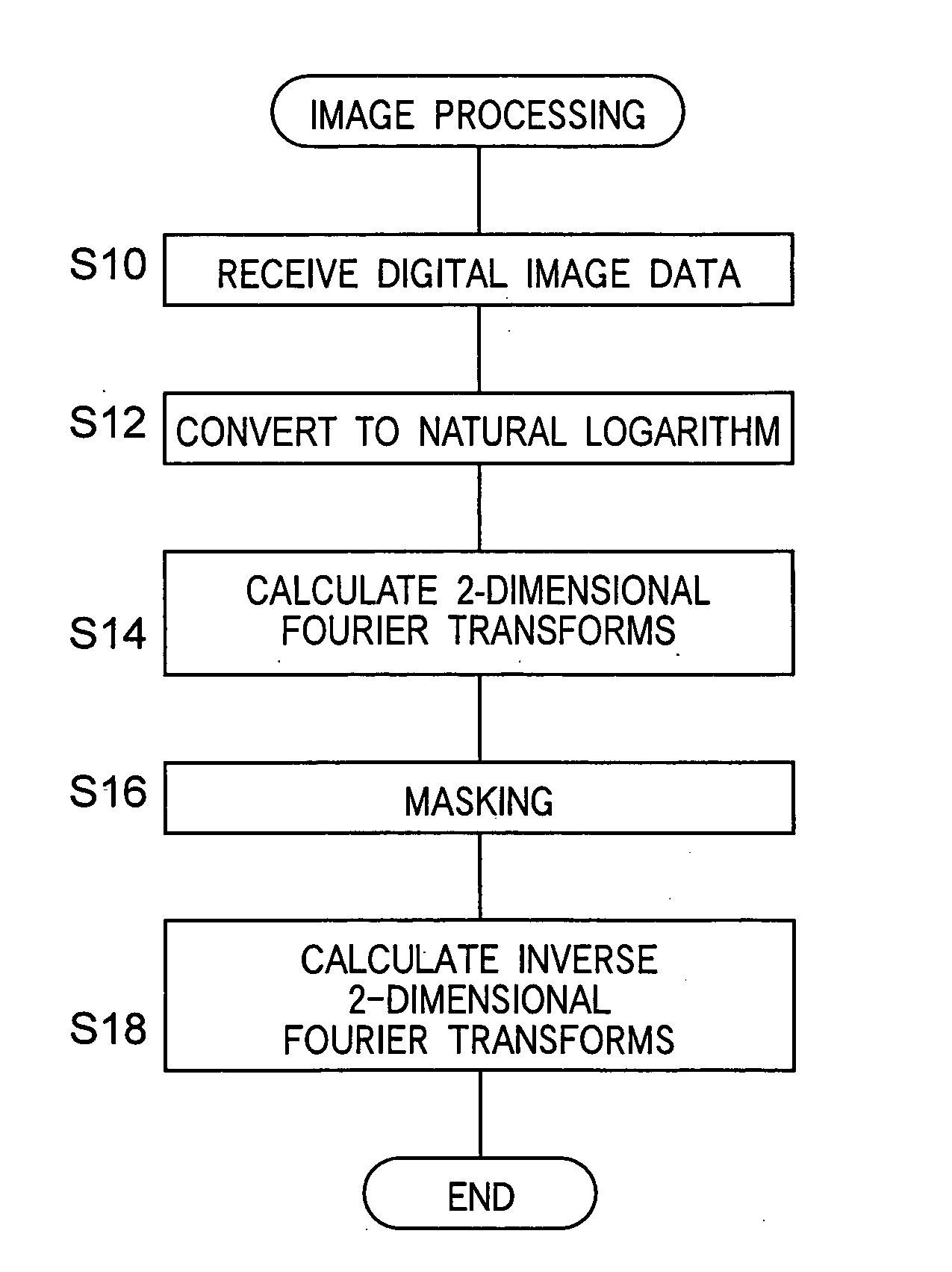
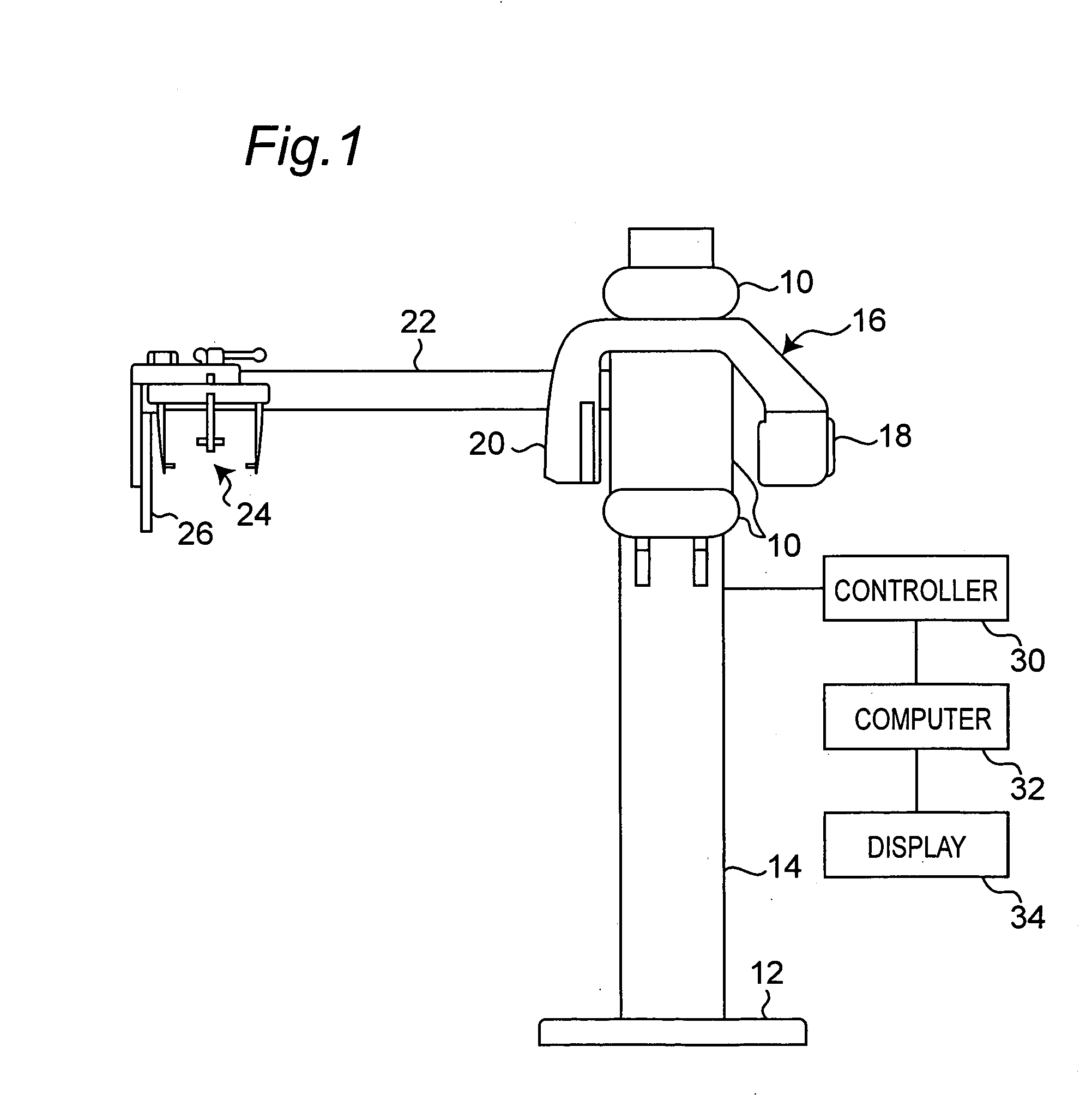
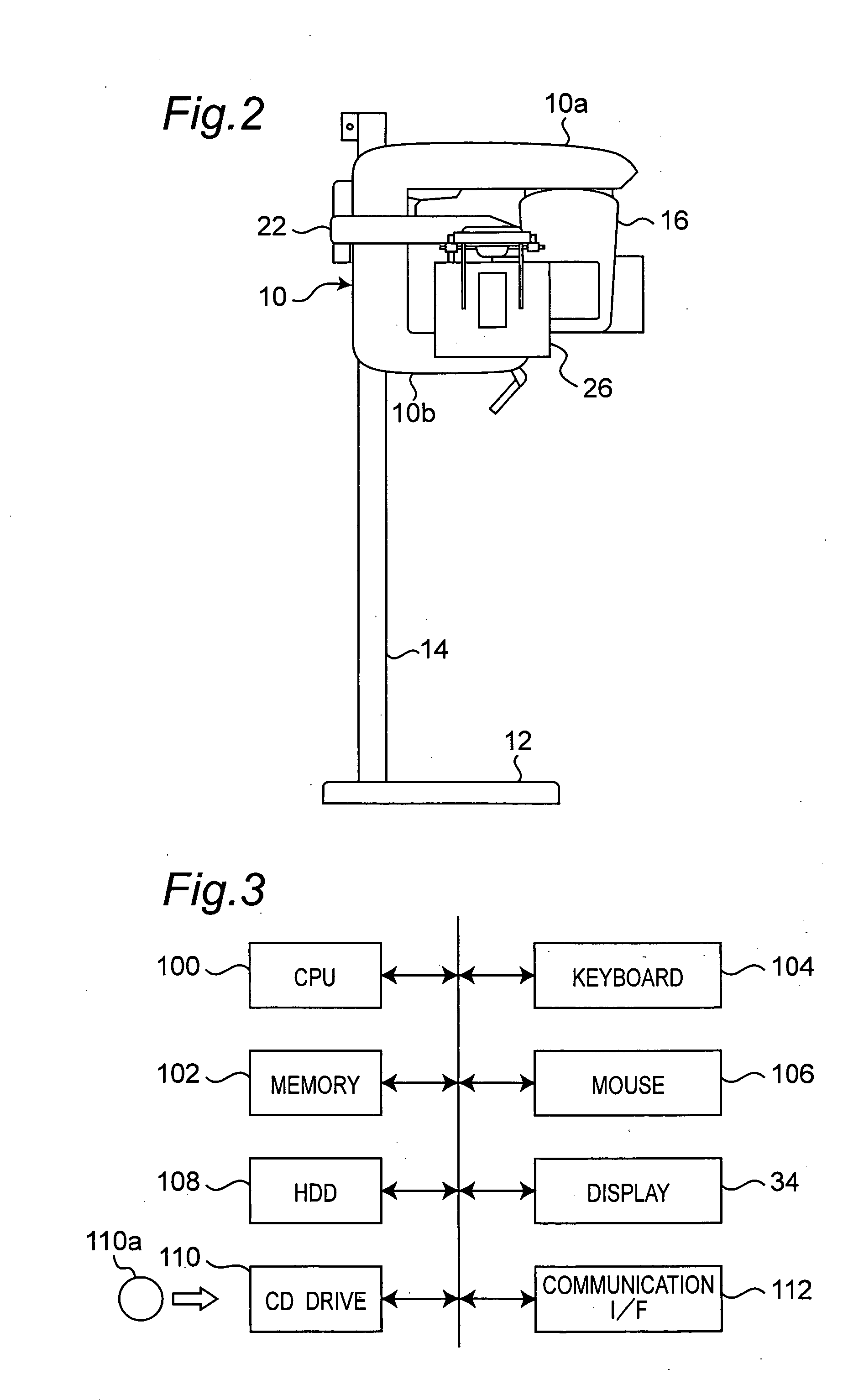
[0021] Referring now to the drawings, wherein like reference characters designate like or corresponding parts throughout the views, FIGS. 1 and 2 show an X-ray apparatus used for dental panoramic and cephalo-metric radiography. In this apparatus, a main body 10 of a lift has a central part in parallel to an upright support 14 fixed to a base 12 and upper and lower extensions 10a and 10b extending from the top and from the bottom of the central part towards the front of the apparatus. A lifting mechanism (not shown) is connected to the central part for moving the main body 10 up or down along the support 14. The upper extension 10a includes therein a device (not shown) positioning a patient. A rotary arm 16 is supported rotatably below the upper extension 10a. The rotary arm 16 has an X-ray head (X-ray source) 18 which generates X-rays and an X-ray sensor 20, such as a film, an imaging plate, a charge-coupled device (CCD) sensor, a metal-oxide-semiconductor (MOS) sensor or an X-ray f...


PUM


Abstract

In a method for processing a digital X-ray image data obtained by panoramic or cephalo-metric radiography, Fourier transforms are computed in a two-dimensional frequency space on the digital X-ray image data, and data of the Fourier transforms are multiplied by values of a mask having different frequency characteristics in two coordinate directions in the two-dimensional frequency space, the values being smaller than one around an origin in the frequency space. Then, inverse Fourier transforms are computed on the data of the Fourier transforms after the multiplication. A resultant image data in real space is provided for diagnosis.

Description

FIELD OF THE INVENTION [0001] The invention relates to image processing of digital X-ray radiographs obtained in panoramic or cephalo-metric radiography. RELATED ART [0002] Panoramic or cephalo-metric radiography is used in dental X-ray radiography. In panoramic radiography, an entire dentition or jawbone and its neighborhood are imaged in a single radiograph. For example, a slit-like X-ray beam generated by an X-ray source scans an object (or a patient) successively while moving a film in synchronization with the scan. Then an image of a slice plane is obtained. In cephalo-metric radiography, a head of a person or an object is fixed to keep a constant position relationship between the X-ray source and the object, and an entire object is imaged in a front view, a side view or the like. [0003] The invention relates to image processing of digital X-ray radiograph. An example of a background art for such image processing is described in Japanese Utility Model laid open Publication 6-31...

Claims


Application Information

Patent Timeline
IPC IPC(8): A61B6/14A61BA61B6/00G06K9/00G06K9/36G06T5/10G06T7/00G06T7/40H04N5/232H04N5/32H04N5/325
CPCG06T5/10G06T2207/30008G06T2207/30036G06T2207/20056G06T5/007G06T2207/10116H04N5/32G06T5/90H04N23/30
Inventor SUZUKIHAYASHI, TAKESHI
Owner MORITA MFG CO LTD
Why Eureka
Eureka Blog
Learn More