
System and method for endoscopic path planning

Inactive Publication Date: 2005-05-19
SIEMENS MEDICAL SOLUTIONS USA INC
View PDF14 Cites 197 Cited by
  • Summary
  • Abstract
  • Description
  • Claims
  • Application Information

AI Technical Summary

Problems solved by technology

Until recently, however, TBBx has been limited to central nodules located close to major airways due to the size of the bronchoscope.
It is difficult, however, to plan a path through numerous branching airways to reach desired peripheral nodules.
Even when using multi-slice computed tomography (CT) imaging systems capable of obtaining image volumes with an isotropic resolution of, for example, 0.5 mm, peripheral airways near the desired nodule may not be visible in the image data due to partial volume effects.
Further, as airways branch repeatedly into smaller peripheral airways, the number of possible pathways increases exponentially, making it difficult to plan a pathway to a desired peripheral nodule.
However, virtual endoscopic techniques are limited by the resolution of the available 3D data, and are thus confined to central airways of the lung that are visible in the CT image data.

Method used


Image

  • System and method for endoscopic path planning
  • System and method for endoscopic path planning
  • System and method for endoscopic path planning

Examples


Embodiment Construction

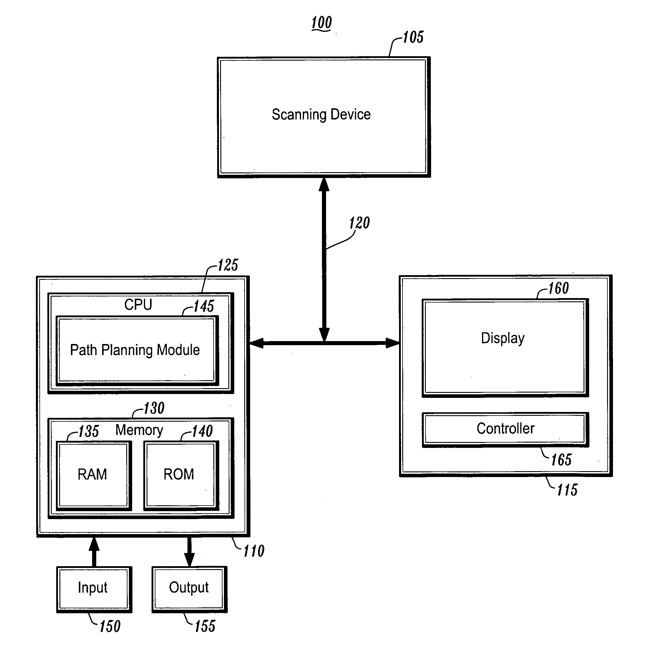
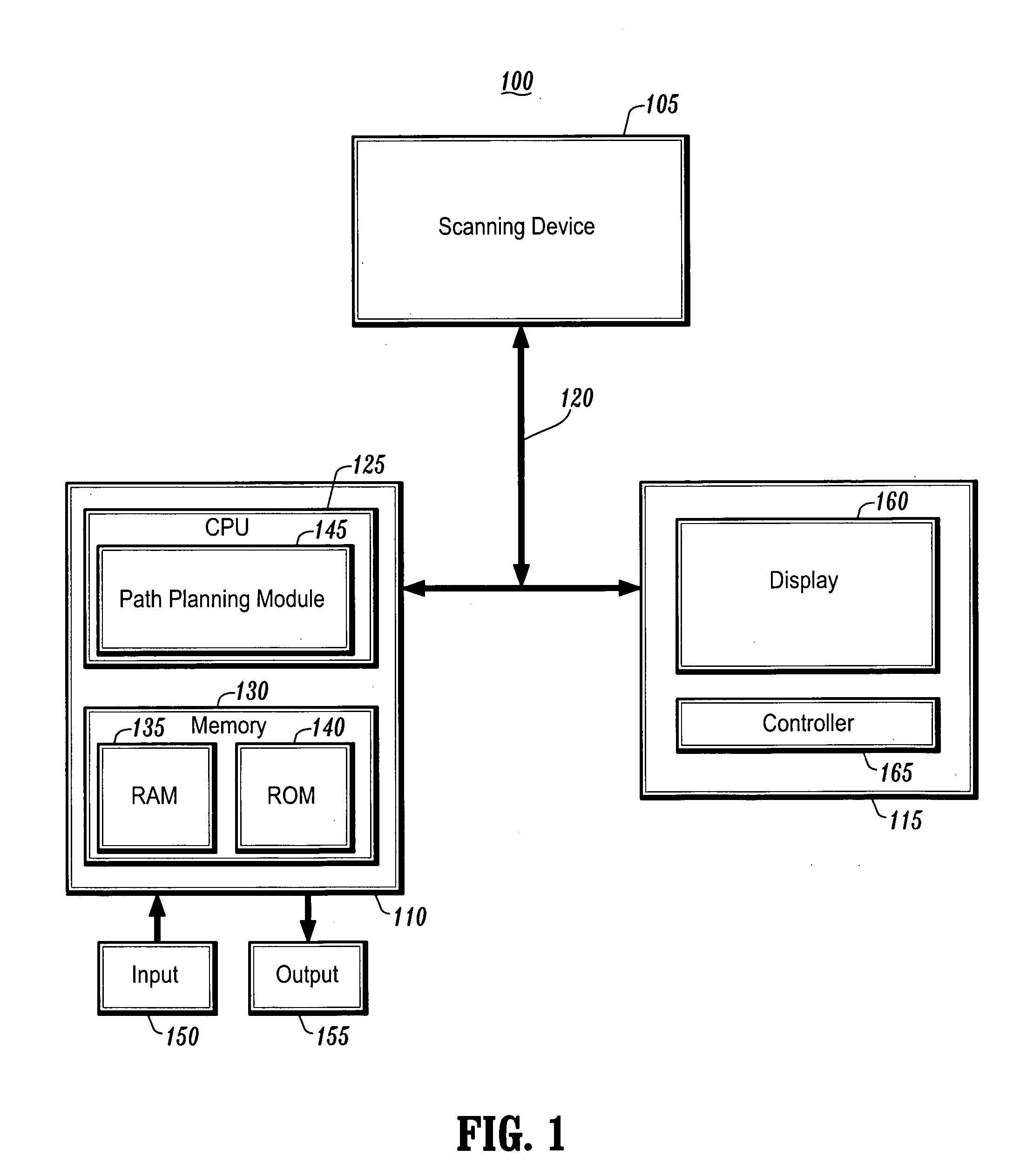
[0037]FIG. 1 is a block diagram of a system 100 for endoscopic path planning according to an exemplary embodiment of the present invention. As shown in FIG. 1, the system 100 includes, inter alia, a scanning device 105, a personal computer (PC) 110 and an operator's console and / or virtual navigation terminal 115 connected over, for example, an Ethernet network 120. The scanning device 105 may be a magnetic resonance imaging (MRI) device, a computed tomography (CT) imaging device, a helical CT device, a positron emission tomography (PET) device, a two-dimensional (2D) or three-dimensional (3D) fluoroscopic imaging device, a 2D, 3D, or four-dimensional (4D) ultrasound imaging device, or an x-ray device, etc.

[0038] The PC 110, which may be a portable or laptop computer, a personal digital assistant (PDA), etc., includes a central processing unit (CPU) 125 and a memory 130, which are connected to an input 150 and an output 155. The CPU 125 includes a path planning module 145 that inclu...


PUM


Abstract

A system and method for endoscopic path planning is provided. The method comprises: identifying a target in a lung, wherein the target is located in a peripheral airway of the lung; generating an endoscopic path to the target, wherein a peripheral artery is used as a surrogate for the peripheral airway; and viewing the endoscopic path.

Description

CROSS-REFERENCE TO RELATED APPLICATIONS [0001] This application claims the benefit of U.S. Provisional Application No. 60 / 486,319, filed Jul. 11, 2003, a copy of which is herein incorporated by reference.BACKGROUND OF THE INVENTION [0002] 1. Technical Field [0003] The present invention relates to endoscopic path planning, and more particularly, to endoscopic path planning using arteries as a surrogate for airways. [0004] 2. Discussion of the Related Art [0005] Bronchoscopy with transbronchial biopsy (TBBx) is a diagnostic procedure in which a bronchoscope is inserted through the nose or mouth to collect multiple specimens of lung tissue. TBBx has been shown to be less traumatic to patients as compared to other methods of biopsying suspicious lung nodules, such as transthoracic needle biopsy (TTNBx) and video assisted thorascopic surgery (VATS). Until recently, however, TBBx has been limited to central nodules located close to major airways due to the size of the bronchoscope. [0006]...

Claims


Application Information

Patent Timeline
IPC IPC(8): A61B5/05A61B19/00G06T7/00G06T17/40
CPCA61B19/50A61B19/52G06T2210/41G06T19/003A61B2019/5236A61B34/10A61B90/36A61B2090/374
Inventor GEIGER, BERNHARDKIRALY, ATILLA PETERNAIDICH, DAVID P.NOVAK, CAROL L.
Owner SIEMENS MEDICAL SOLUTIONS USA INC
Features
  • Generate Ideas
  • Intellectual Property
  • Life Sciences
  • Materials
  • Tech Scout
  • Unparalleled Data Quality
  • Higher Quality Content
  • 60% Fewer Hallucinations
Learn More