Methods for detecting anionic and non-anionic compositions using carbocyanine dyes
a technology of carbocyanine dye and anionic composition, which is applied in the field of methods for detecting anionic and nonanionic compositions using carbocyanine dye, can solve the problems of large amount of protein, limited linear dynamic range, and difficult protein quantitation
- Summary
- Abstract
- Description
- Claims
- Application Information
AI Technical Summary
Problems solved by technology
Method used
Image

Examples
example 1
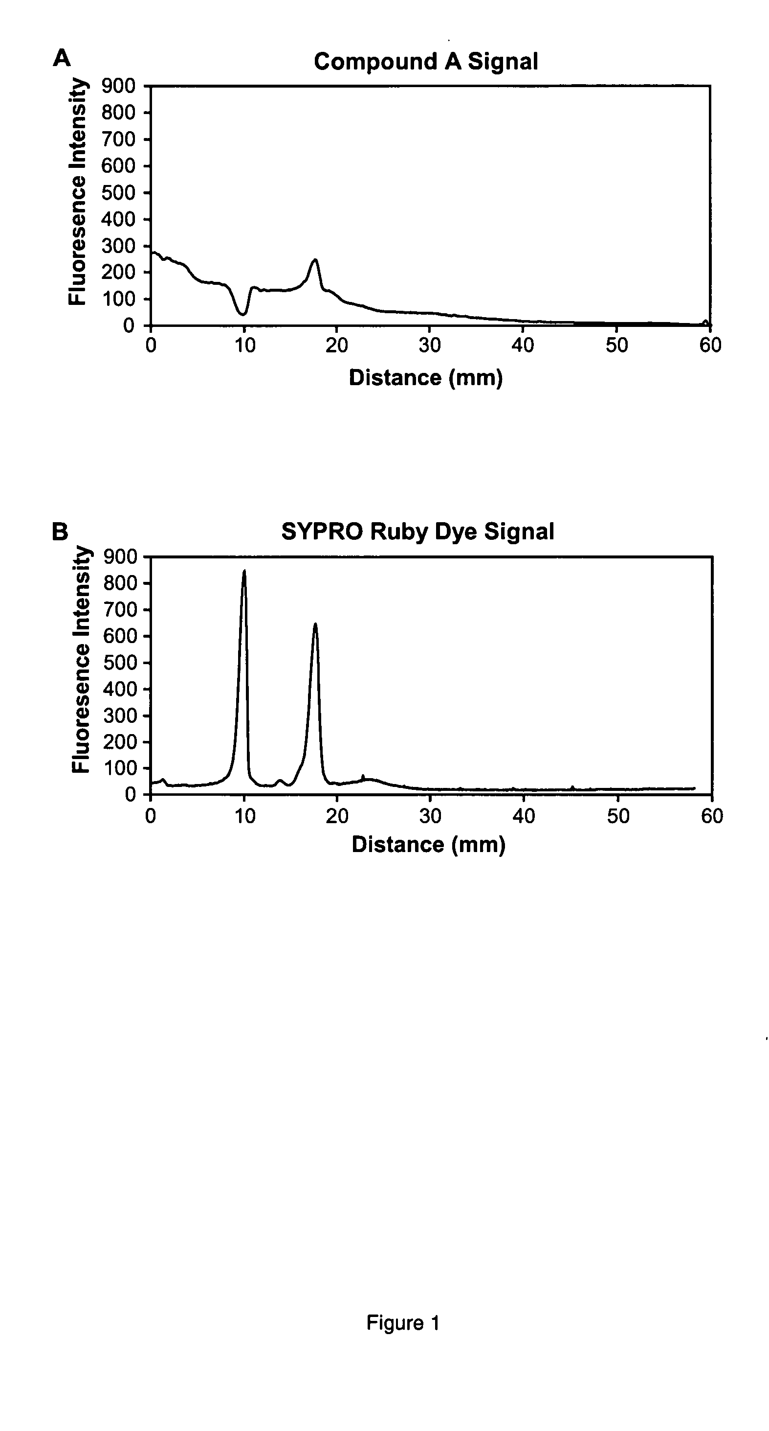
Serial Dichromatic Detection of Proteins in SDS-Polyacrylamide Gels Using (A) and SYPRO Ruby Protein Gel Stain
[0187] Proteins were separated by SDS-polyacrylamide gel electrophoresis utilizing 13% T, 2.6% C gels. % T is the total monomer concentration expressed in grams per 100 mL and % C is the percentage crosslinker. The 0.75 mm thick, 6×10 cm gels were subjected to electrophoresis using the Bio-Rad mini-Protean III system according to standard procedures. Following separation of the proteins on SDS-polyacrylamide gels, the gels were fixed for one hour in 100 mL of 50% ethanol / 7% acetic acid and then fixed overnight in 100 mL of fresh fixative solution to ensure complete elimination of SDS. Gels were next washed 3 times for 20 min each in deionized water. The gels were then incubated in a staining solution containing 4 μM (A), 10% ethanol, 20 mM MOPS at pH 7.25 for 2 h in a total volume of 50 mL. Afterwards, the gels were washed for 30 min in 50 mL of 20% acetonitrile, 10 mM M...
example 2
Selectivity of Staining of the Phosphoprotein Chicken Ovalbumin Relative to the Nonphosphorylated Protein Bovine Serum Albumin in SDS Polyacrylamide Gel Electrophoresis
[0189] A mixture containing 500 ng each of the purified proteins chicken ovalbumin and bovine serum albumin was prepared in 1×SDS sample buffer (50 mM Tris, 10% glycerol, 50 mM DTT, 2% SDS, and 0.01% bromophenol blue, pH 6.8). Proteins were separated by SDS-polyacrylamide gel electrophoresis, stained with (A), imaged, stained for total proteins with SYPRO Ruby dye, and imaged again as described in Example 1. Bovine serum albumin was negatively stained by (A) aggregate, such that the signal was less than the background signal of (A) aggregate, obtained from a blank region of the gel. Chicken ovalbumin was positively stained by (A) aggregate, such that the signal was higher than the background signal of unbound (A) aggregate. Post-staining of the gel with SYPRO Ruby dye shows two peaks of approximately equal intensity...
example 3
Selective Staining of Anionic Proteins in SDS Polyacrylamide Gel Electrophoresis
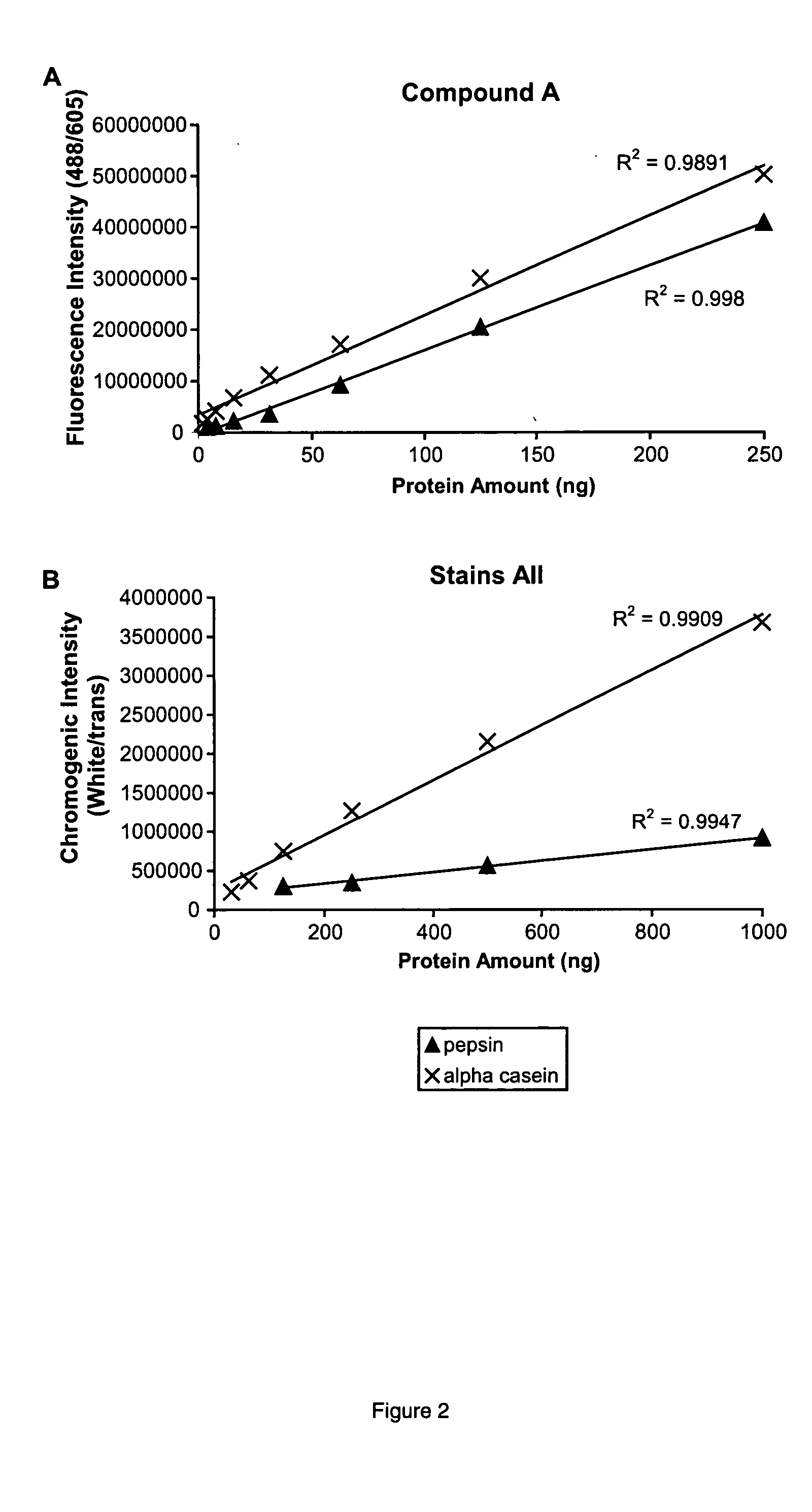
[0190] Samples containing 500 ng of pepsin and α casein (both phosphoproteins), chicken ovalbumin (a phosphorylated glycoprotein), Tamm-Horsfall glycoprotein, (contains numerous sulfated glycans), α1 acid glycoprotein (an acidic glycoprotein), avidin (a basic glycoprotein), and the proteins phosphorylase b, bovine serum albumin, carbonic anhydrase, and lysozyme were prepared in 1×SDS sample buffer. Proteins were separated by SDS-polyacrylamide gel electrophoresis, stained with (A), imaged, stained for total proteins with SYPRO Ruby dye, and imaged again as described in Example 1. The (A) aggregate stained the acidic phosphoprotein, pepsin, the most strongly of the proteins in the sample, followed by α casein, at 79% of the intensity of pepsin. The (A) aggregate stained α1 acid glycoprotein, Tamm-Horsfall glycoprotein, and chicken ovalbumin at 29%, 12%, and 3% relative to pepsin. Phosphorylase b, bovine...
PUM
 Abstract
Description
Claims
Application Information
 - R&D Engineer
- R&D Manager
- IP Professional
- Industry Leading Data Capabilities
- Powerful AI technology
- Patent DNA Extraction
Browse by: Latest US Patents, China's latest patents, Technical Efficacy Thesaurus, Application Domain, Technology Topic.
© 2024 PatSnap. All rights reserved.Legal|Privacy policy|Modern Slavery Act Transparency Statement|Sitemap