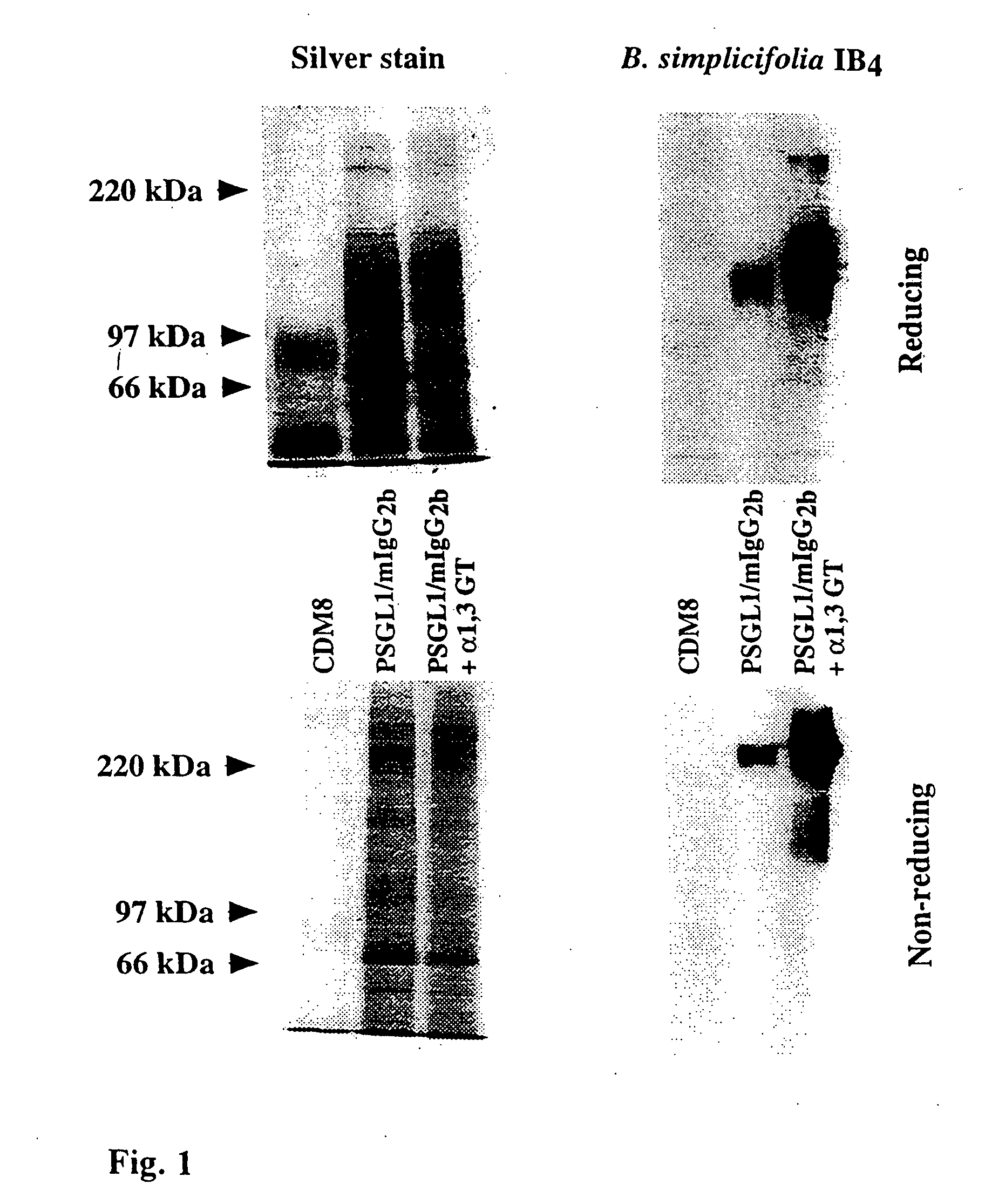
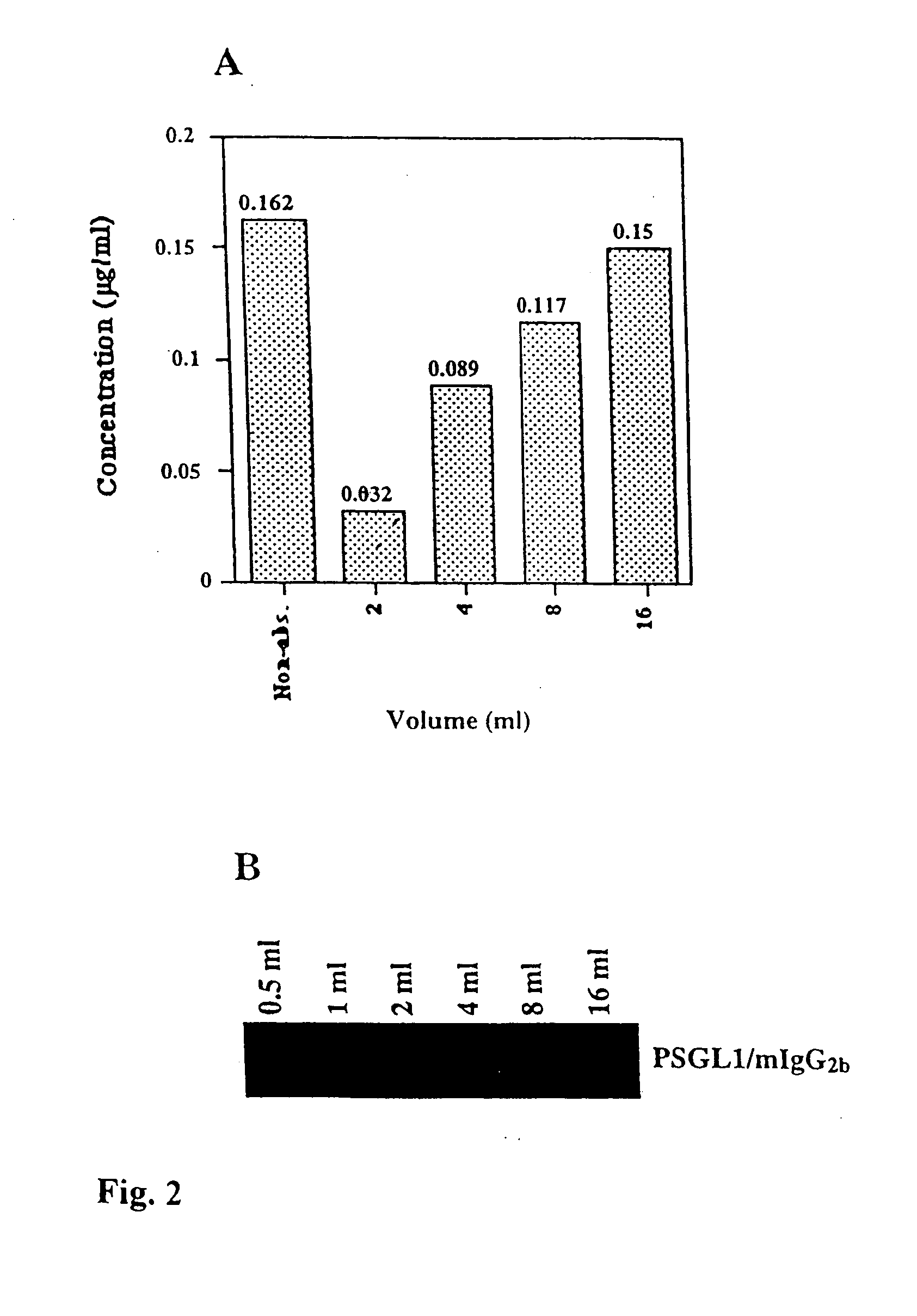
[0018] Thus, as the fusion protein according to the present invention may be prepared by the culture of genetically manipulated cells, such as COS cells, it is both cheaper and easier to produce than the previously saccharides produced by organic synthesis. Even though a COS cell expressing the Galα1,3Gal epitope on its surface has been described (12), the present invention is the first proposal of a recombinant fusion protein, which carries multiple such epitopes. The fusion protein according to the invention can easily be designed to include other peptides and parts, which may be advantageous for a particular application. Examples of other components of the fusion protein according to the invention will be described more detailed below.
[0019] Thus, in a preferred embodiment, the antigenic fusion protein according to the invention further comprises a part, which mediates binding to selectin, such as P-selectin. Said part is preferably a highly glycosylated protein, such as a protein of mucin type. The mucins are due to their high content of O-linked carbohydrates especially advantageous together with the Galα1,3Gal epitope in the fusion protein according to the invention, as the antigenic properties in the present context thereby are greatly improved. Thus, it has been shown that the binding of antibodies which are reactive with the Galα1,3Gal epitope is even more efficient if said epitope is presented by a protein of mucin type, which indicates that the binding in fact may involve more than said epitope alone.
[0027] Yet another aspect of the present invention is a vector, which comprises the cDNA molecule as described above together with appropriate control sequences, such as primers etc. The skilled in the art can easily choose appropriate elements for this end.
[0028] Another aspect of the present invention is a cell line transfected with the above defined vector. The cell line is preferably eukaryotic, e.g. a COS cell line. In the preferred embodiment said cells are secreting the fusion protein according to the invention into the culture medium, which makes the recovery thereof easier and, accordingly, cheaper than corresponding methods for synthesis thereof would be.
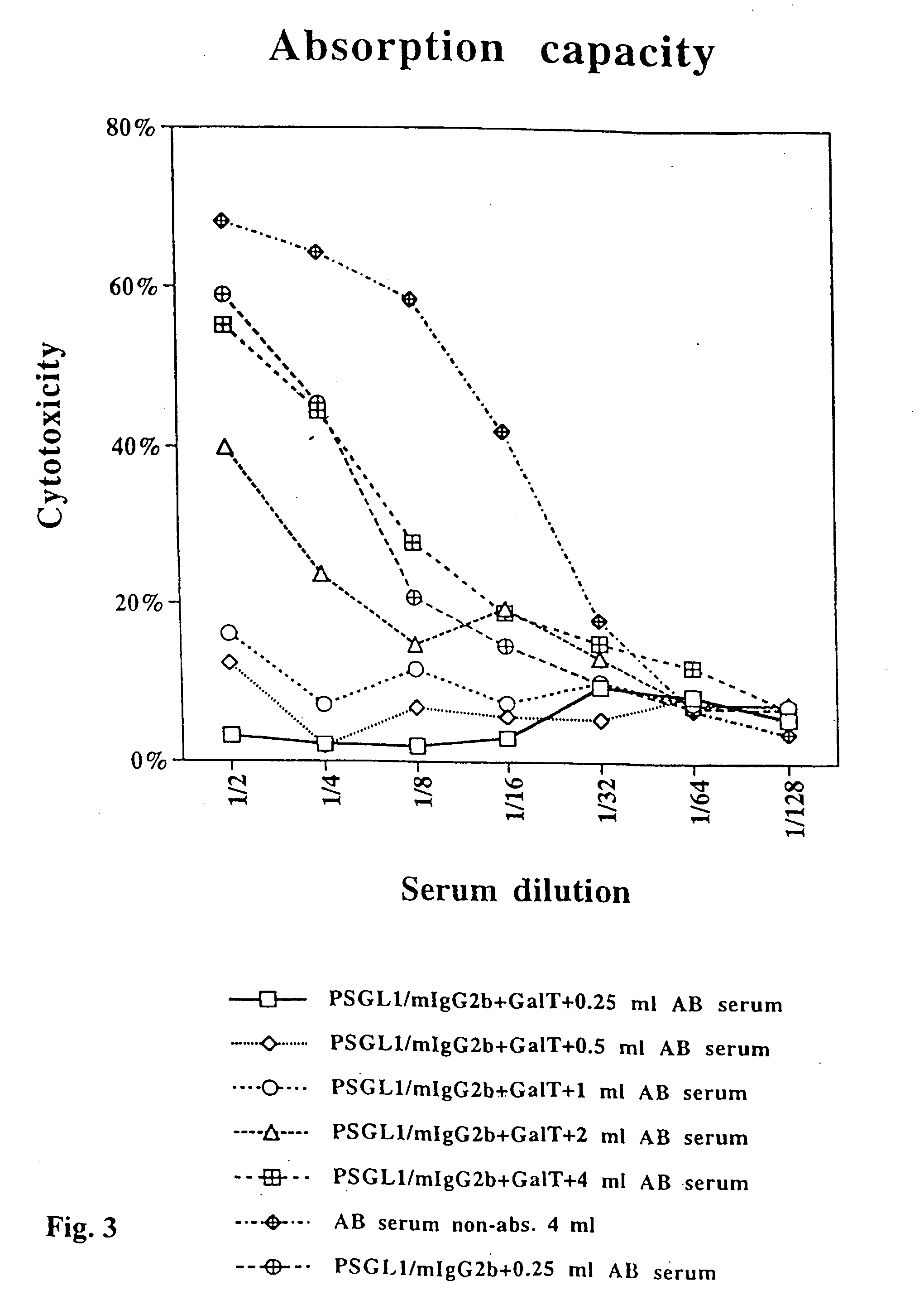
[0046] The absorption capacity of immobilized, Galα1,3Gal-substituted PSGL1 / mIgG2b. Twenty ml of supernatant from COS cells transfected with the PSGL1 / mIgG2b plasmid alone or together with the porcine α1,3GT cDNA, were mixed with 500 μl gel slurry of anti-mouse IgG agarose beads each. Following extensive washing the beads were aliquoted such that 100 μl of gel slurry (50 μl packed beads) were mixed with 0.25, 0.5, 1.0, 2.0 and 4.0 ml of pooled, complement-depleted, human AB serum, and rolled head over tail at 4° C. for 4 hrs. Following absorption on PSGL1 / mIgG2b and Galα1,3Gal-substituted PSGL1 / mIgG2b, the serum was assayed for porcine endothelial cell cytotoxicity in the presence of rabbit complement using a 4 hr 51Cr release assay (FIG. 3). As shown in FIG. 3, 100 μl of beads carrying approximately 300 ng of PSGL1 / mIgG2b (see above) can reduce the cytotoxicity of 4 and 2 ml AB serum in each dilution step, and completely absorb the cytotoxicity present in 1 ml and less of human AB serum. Note that the same amount of non-Galα1,3Gal-substituted PSGL1 / mIgG2b reduces the cytotoxicity of 0.25 ml absorbed human AB serum only slightly (FIG. 3).
[0048] The effect of Galα1,3Gal-substituted PSGL1 / mIgG2b on ADCC of porcine endothelial cells. Several assays have been performed under serum-free conditions where PEC-A have had an intermediate sensitivity to direkt killing by freshly isolated PBMC when compared to K562 and Raji cells; K562 being sensitive and Raji non-sensitive to killing by human NK cells (FIG. 6A). In the presence of 10% human, complement-inactivated AB serum, the killing rate was almost doubled supporting an ADCC effect in vitro (FIG. 6B) in agreement with previously published data (30). However, if the serum is absorbed with the Galα1,3Gal-substituted PSGL1 / mIgG2b under conditions known to remove all PEC-A cytotoxic antibodies (se above), the killing rate decreases to levels slightly lower than those seen under serum-free conditions. On the other hand, the PSGL1 / mIgG2b fusion protein itself without Galα1,3Gal epitopes could not absorb out what caused the increased killing rate in the presence of human AB serum (FIG. 6B). These data support the notion that anti-pig antibodies with Galα1,3Gal specificity can support an antibody-dependent cell-mediated cytotoxicity in vitro, and that the Galα1,3Gal-substituted PSGL1 / mIgG2b fusion protein can effectively remove these antibodies just as it effectively removes the complement-fixing cytotoxic anti-pig antibodies.


