Image generation apparatus, image generation method, and program therefor

a technology of image generation and image, applied in the field of image generation apparatus, image generation method, and program therefor, can solve the problems of inaccurate detection of abnormalities, no image, and inability to accurately carry out positional matching, so as to facilitate the detection of abnormalities, the effect of more effectiveness and accuracy

Active Publication Date: 2006-01-05
FUJIFILM CORP +1
View PDF5 Cites 22 Cited by
  • Summary
  • Abstract
  • Description
  • Claims
  • Application Information

AI Technical Summary

Benefits of technology

[0012] The present invention has been conceived based on consideration of the above circumstances. An object of the present invention is therefore to provide an image generation method, an image generation apparatus, and a program for enabling detection of an abnormality with more effectiveness and accuracy than conventional image reading by comparison.
[0028] The image generation apparatus of the present invention may further comprise abnormality detection means for detecting a candidate area of an abnormality from the medical image so that the artificial image generation means can generate the artificial image corresponding to the segment including a position at which the candidate area has been detected.
[0035] According to the present invention, the divided segments are classified according to the similar anatomic characteristics represented by the structures in the medical image generated by radiography of the predetermined part of the subject such as a human body, and the artificial image can be generated for each of the classified segments. Therefore, the artificial image is generated according to the characteristics of each of the segments. In addition, the artificial image can be generated regarding only the segment having been specified, instead of the entire chest, which is efficient.
[0036] If the abnormality candidate area is detected and if the artificial image is generated for the segment including the position of the abnormality candidate area, the image of only the part corresponding to the abnormality is generated for image reading by comparison. In this manner, efficiency and detection accuracy can be improved.
[0039] In the case where the medical image is a chest image obtained by radiography of a chest, if the segments can be classified according to the overlap of the ribs and the positions in the lung fields based on recognition of lung fields and ribs, detection accuracy can be improved when an abnormality such as that of lung cancer is diagnosed.

Problems solved by technology

Therefore, in the case where a large three-dimensional positional difference (leaning forward or backward or twisting sideways) is observed due to a posture change of the patient in images, positional matching is not carried out accurately.
Therefore, an artifact is generated in a subtraction image, which leads to inaccurate detection of an abnormality.
Furthermore, in the case where a patient does not have any image radiographed in the past, no image is used for comparison.
Therefore, the methods using a subtraction image cannot be used, regardless of effectiveness of the methods for detecting an abnormal pattern such as that indicative of lung cancer, which tends to be missed.
Therefore, generating an artificial image of the entire chest is not efficient.

Method used


Image

  • Image generation apparatus, image generation method, and program therefor
  • Image generation apparatus, image generation method, and program therefor
  • Image generation apparatus, image generation method, and program therefor

Examples


Embodiment Construction

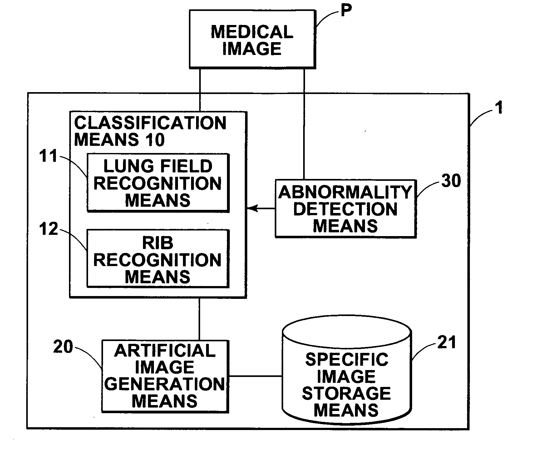
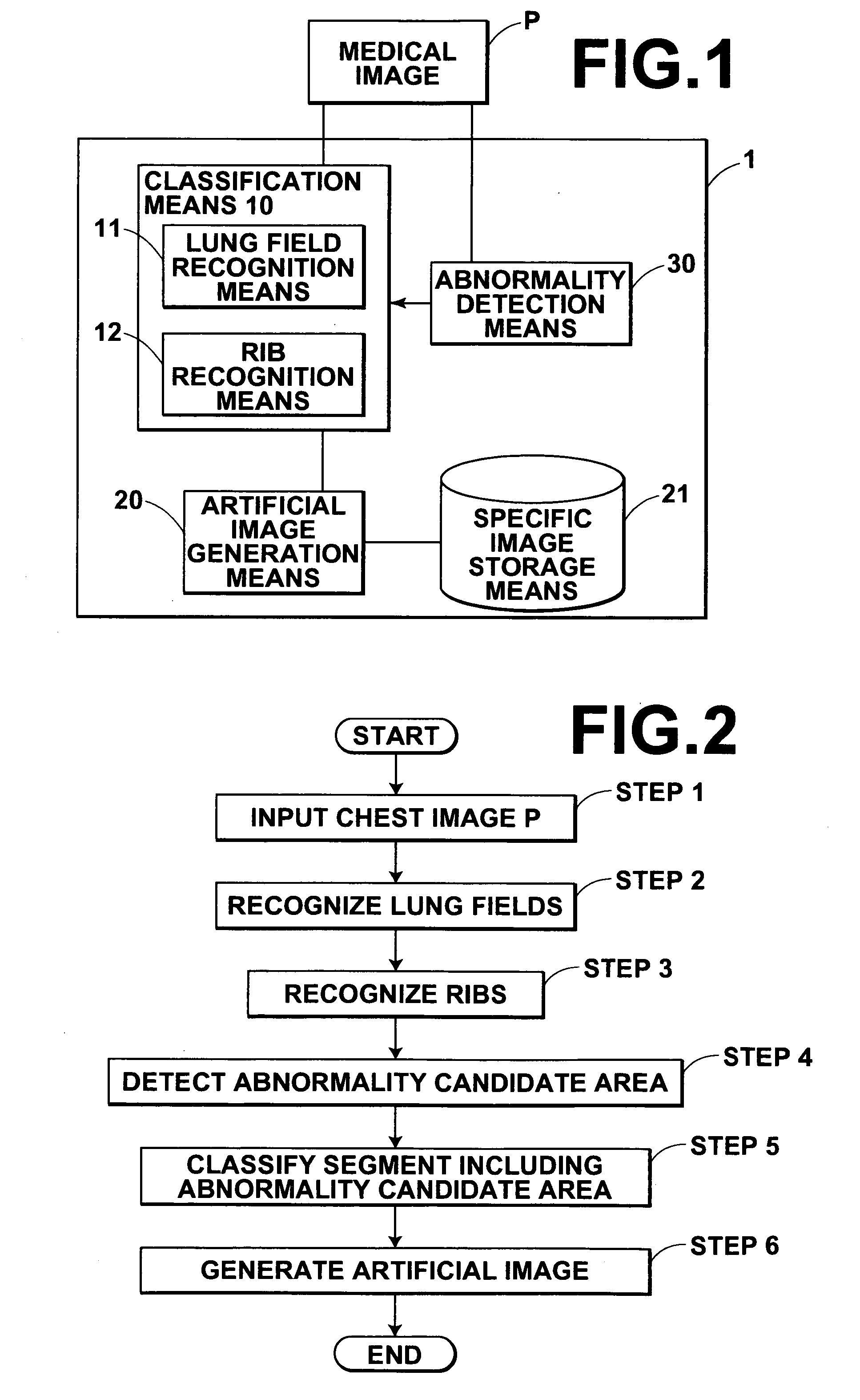
[0059] Hereinafter, an embodiment of an image generation apparatus of the present invention will be described with reference to the accompanying drawings. As shown in FIG. 1, an image generation apparatus 1 in this embodiment comprises classification means 10 for classifying segments obtained by division of a medical image P according to similar anatomic characteristics, abnormality detection means 30 for detecting a candidate area of an abnormality from the image, and an artificial image generation means 20 for generating an artificial image representing normal structures of a segment corresponding to one of the segments including the abnormality candidate area that has been detected.

[0060] The artificial image generation means 20 has Eigen-image storage means 21 for storing Eigen-images each representing specific characteristics of normal structures in each of the segments having been classified according to the anatomic characteristics. The artificial image generation means 20 g...


PUM


Abstract

An artificial image used for comparison with a medical image can be generated efficiently and accurately. A medical image including anatomically characteristic structures obtained by radiography of a predetermined part of a subject such as a human body is classified into segments having similar anatomic characteristics represented by the structures. The artificial image is generated for a specified one of the segments regarding the structures in normality.

Description

BACKGROUND OF THE INVENTION [0001] 1. Field of the Invention [0002] The present invention relates to an image generation apparatus and an image generation method for aiding detection of an abnormality candidate by reading a medical image. The present invention also relates to a program that causes a computer to execute the image generation method. [0003] 2. Description of the Related Art [0004] In the field of medicine, two or more medical images of the same patient radiographed at different times have been compared for detecting an abnormality based on a difference between the images and for discussing therapeutic measures by understanding the progression or remission of a disease, for example. [0005] A small abnormal pattern such as that indicative of lung cancer at an early stage is especially easy to miss. Therefore, Japanese Unexamined Patent Publication No. 2002-158923, for example, describes a method for detecting a candidate of such an abnormality. In this method, subtractio...

Claims


Application Information

Patent Timeline
Patent Type & Authority Applications(United States)
IPC IPC(8): A61B5/05
CPCG06K9/4609G06K9/6247G06T2207/30061G06T7/0012G06K2209/05G06V10/443G06V2201/03G06V10/7715G06F18/2135
Inventor OOSAWA, AKIRA
Owner FUJIFILM CORP
Why Eureka
Try Eureka