Systems, methods and apparatus for dual mammography image detection

a mammography and image detection technology, applied in the field of mammography imaging system, can solve the problems of duplicate hardware, inability to provide mammogram, and inability to accurately detect breast tissue x-rays,

Inactive Publication Date: 2006-04-06
GENERAL ELECTRIC CO
View PDF8 Cites 51 Cited by
  • Summary
  • Abstract
  • Description
  • Claims
  • Application Information

AI Technical Summary

Problems solved by technology

However, X-ray imaging of breast tissue has the inherent limitation in that a mammogram provides only a planar image of a three-dimensional object.
However, much of the hardware, such as computer and display, are duplicated because the systems are built and sold separately.

Method used


Image

  • Systems, methods and apparatus for dual mammography image detection
  • Systems, methods and apparatus for dual mammography image detection
  • Systems, methods and apparatus for dual mammography image detection

Examples


an embodiment

Methods of an Embodiment

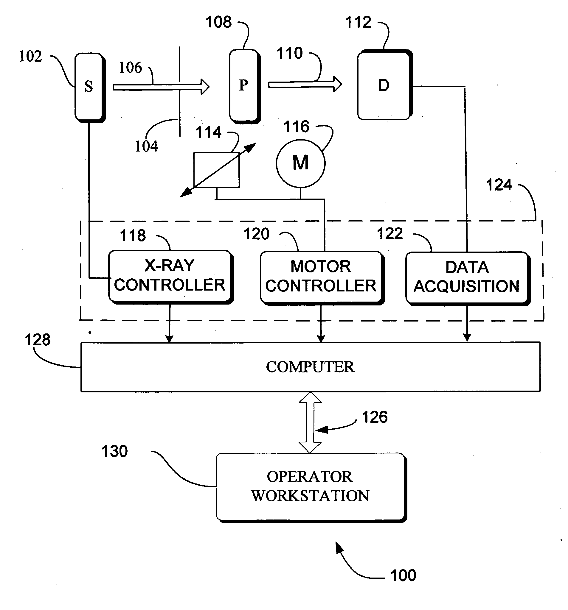
[0059] In the previous section, a system level overview of the operation of an embodiment was described. In this section, the particular methods performed by the server and the clients 128 and 130 of such an embodiment are described by reference to a series of flowcharts. Describing the methods by reference to a flowchart enables one skilled in the art to develop such programs, firmware, or hardware, including such instructions to carry out the methods on suitable computerized clients the processor of the clients executing the instructions from computer-readable media. Similarly, the methods performed by the server computer programs, firmware, or hardware are also composed of computer-executable instructions. Methods 1100-150000 are performed by a client program executing on, or performed by firmware or hardware that is a part of a computer, a microprocessor, or controller and is inclusive of the acts required to be taken by the computer 128 or workstation 13...


PUM


Abstract

Systems and methods are provided by which a mammography imaging system offers X-ray and ultrasound imaging that allows sharing of common hardware such as the computer and display. Small regions of interest are imaged with X-ray at higher image quality by using a second sensor with higher DQE than the full-field sensor can obtain. In some embodiments a specialized chamber is provided for securing the anatomy to a fixed location, ultrasound image data is collected along with ultrasound probe location and orientation data from sensors on a handheld probe from which data images can be viewed directly, or used to reconstruct tomographic images of any desired cross-section, or used for various “3-D” image visualization methods. An imaging schedule defined by location and orientation of an ultrasound probe is used to generate a three-dimensional ultrasound image.

Description

FIELD OF THE INVENTION [0001] This invention relates generally to mammography imaging system, and more particularly to higher detective quantum efficiency images. BACKGROUND OF THE INVENTION [0002] The use of X-ray technology for providing two-dimensional images of breast tissue for diagnosis of carcinoma or other abnormalities is in wide use. However, X-ray imaging of breast tissue has the inherent limitation in that a mammogram provides only a planar image of a three-dimensional object. [0003] The detective quantum efficiency (“DQE”) of an image is the conventional measure of X-ray image quality. In simpler terms, the DQE is the resolution of the detector. DQE is constant across an image for a given detector and dose technique. [0004] When a potential area of medical concern is indicated on a mammogram, the elevation or depth of the subject area within the two-dimensional image of the breast may be uncertain. Present digital X-ray imagers provide full field or nearly full field im...

Claims


Application Information

Patent Timeline
Patent Type & Authority Applications(United States)
IPC IPC(8): A61B5/05
CPCA61B6/04A61B6/502A61B8/0825A61B8/4209A61B8/58A61B8/483A61B6/4266A61B6/548A61B8/565A61B8/4218
Inventor NEUMANN, DAVID C.VAFI, HABIB
Owner GENERAL ELECTRIC CO
Why Eureka
Try Eureka