Interproximal cavitation detection device and method

a detection device and interproximal cavitation technology, applied in the field of dentistry, can solve the problems of inability to easily recalcify the cavitated surface (remineralized), inability to easily clean the plaque with dental floss, and inability to achieve the effect of interproximal lesions

Inactive Publication Date: 2006-04-20
TEMPLE UNIVERSITY
View PDF12 Cites 8 Cited by
  • Summary
  • Abstract
  • Description
  • Claims
  • Application Information

AI Technical Summary

Benefits of technology

[0007] The present invention is directed to a device and method for diagnosis of enamel cavitation of an interproximal surface that is not readily amendable to probing by instrumentation. More specifically, the device has a substrate of generally rectangular shape and of a sufficient thickness such that it may be inserted into the interproximal space of teeth. Disposed

Problems solved by technology

A cavitated surface cannot be readily re-calcified (remineralized) and cannot be readily cleaned of plaque with dental floss.
This technique will not work for interproximal lesions, i.e., lesions occurring on surfaces of a tooth that are between teeth.
One drawback to implanting spacers is that some patients may refuse to wear them.
Another drawback is that after treatment the teeth must move back together, and this takes time.
Additional drawbacks to implanting spacers include the time required for the spacing to develop, which can take hours to days, the limited access gained due to t

Method used


Image

  • Interproximal cavitation detection device and method
  • Interproximal cavitation detection device and method
  • Interproximal cavitation detection device and method

Examples


example 1

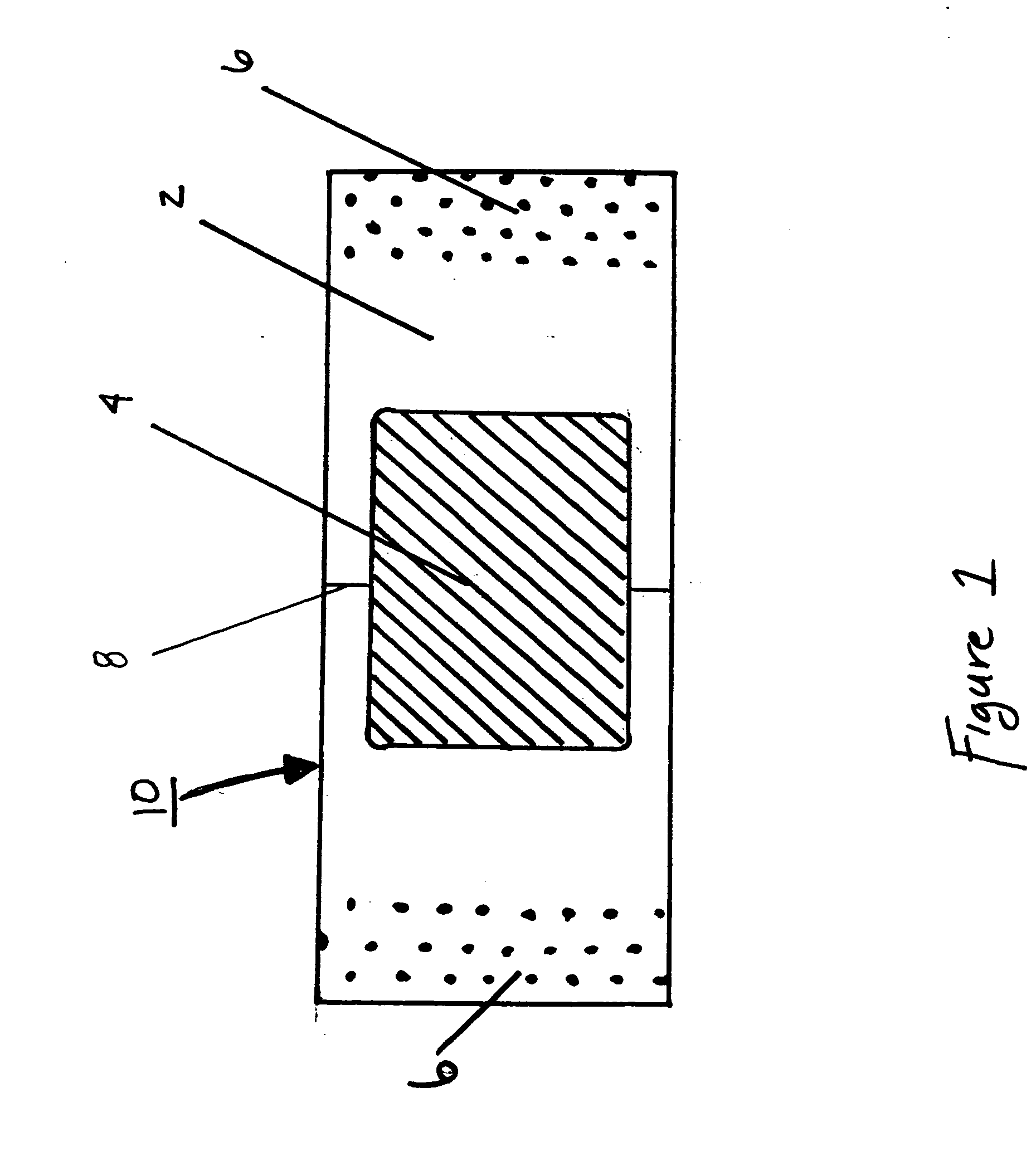
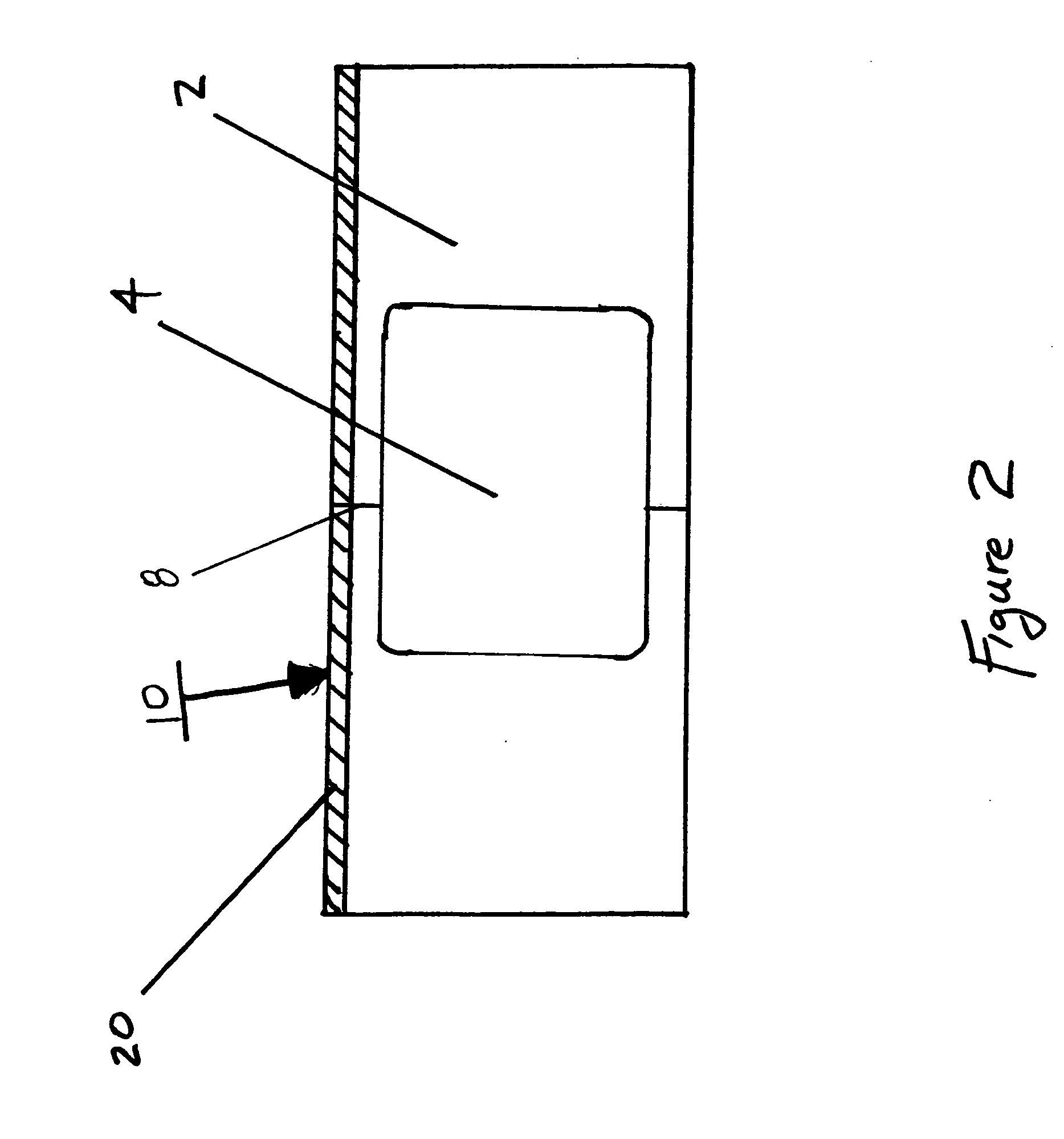
[0032] A Columbia™ typodont artificial teeth and jaw model showing a distal surface with a simulated cavity created with a #4FG bur was used to replicate a human mouth. A polyester fabric strip was cut to size of 7×70 mm to serve as the substrate.

[0033] Polyvinyl putty was applied to the substrate by placing a 0.20 mm thick plastic stencil having a 4×9 mm opening on one edge. The open edge of the stencil was aligned with the edge of the substrate strip. A small portion of the putty was placed on the stencil. With the standard microscope slide held at a sharp angle, the putty was applied to the substrate by dragging the microscope slide across the stencil opening. A resulting 4×9 mm and 0.20 mm impressionable surface was disposed on the substrate.

example 2

[0034] The following is a prophetic exemplary method for detection of a cavitated interproximal surface from a caries lesion.

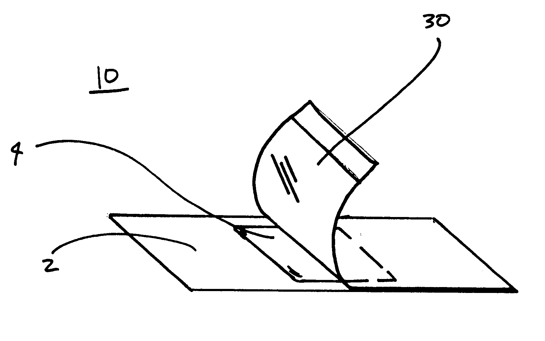
[0035] The teeth in question are professionally cleaned and polished, including flossing, to remove miscellaneous debris that would interfere with the detection process. The interproximal surface of the tooth in question is then air-dried. A sterile promixal cavitation detection device is removed from its packaging.

[0036] With the leading rigid edge, the device is inserted into the interproximal space making sure to avoid premature contact of the impressionable surface against the tooth. The impressionable surface is then aligned with the lesion using a visual indicator by sliding the device laterally thereby bringing the impressionable surface into the interproximal embrasure space and mated therewith. Pressure is applied against the interproximal surface of the tooth with the impressionable material by pulling the ends of the device with equal pressure tow...


PUM


Abstract

The present invention is direct to a device and method for diagnosis of enamel cavitation of an interproximal dental surface. And in particular, the invention is directed to a device and method for diagnosing enamel cavitation of an interproximal tooth surface that is not visible or probe-able by instrumentation in human permanent and deciduous teeth. More specifically, the device has a substrate of generally rectangular shape and of a sufficient thickness such that it may be inserted into the interproximal space of teeth. On the substrate is disposed an impressionable material for contacting the interproximal surface and forming an impression thereof. The impressionable material has visco-elastic properties such that an impression of enamel cavitation on the interproximal surface remains on the impressionable material following removal of the substrate from the interproximal surface.

Description

BACKGROUND OF THE INVENTION [0001] The present invention relates generally to the field of dentistry, and more specifically, to a device and method for diagnosis of enamel cavitation in interproximal carious enamel lesions of teeth. [0002] Enamel carious lesions generally involve decalcification of enamel. Early onset lesions contain macroscopically intact enamel without cavitation on the tooth surface and have the potential for remineralization. Advanced enamel lesions contain a broken, that is, a “cavitated” enamel surface that cannot be repaired by remineralization. Treatment of an advanced lesion thus requires tooth preparation and filling by a trained professional. [0003] The integrity of the outer surface of the tooth is essential for the possibility of remineralization. If the outer surface is intact, it protects the etched inner crystals of hydroxyapatite from being coated by salivary proteins and bacterial plaque. A smooth outer surface also allows the tooth to be cleaned w...

Claims


Application Information

Patent Timeline
IPC IPC(8): A61C5/00A61C9/00
CPCA61B5/1078A61B5/4547A61C19/04
Inventor BOSTON, DANIEL W.
Owner TEMPLE UNIVERSITY
Why Eureka
Try Eureka