Method for evaluation of medical findings in three-dimensional imaging, in particular in mammography

a three-dimensional imaging and medical findings technology, applied in the field of mammography, can solve the problems of difficult evaluation of examination methods by many physicians, difficulty in achieving serial examination of patients with a patient throughput, etc., and achieve the effect of improving the ability to make a medical

Inactive Publication Date: 2006-05-11
SIEMENS AG
View PDF4 Cites 29 Cited by
  • Summary
  • Abstract
  • Description
  • Claims
  • Application Information

AI Technical Summary

Benefits of technology

[0008] An object of the present invention is to improve the ability to make a medical finding in three-dimensional imaging, in particular in mammography.

Problems solved by technology

Such imaging examination methods therefore are not easy for many physicians to evaluate (with regard to the effort expended to make a medical finding) for screening purposes, thus presenting difficulties to accomplish serial examination of patients with a patient throughput of, for example, more than 50 people per day.

Method used


Image

  • Method for evaluation of medical findings in three-dimensional imaging, in particular in mammography
  • Method for evaluation of medical findings in three-dimensional imaging, in particular in mammography
  • Method for evaluation of medical findings in three-dimensional imaging, in particular in mammography

Examples


Embodiment Construction

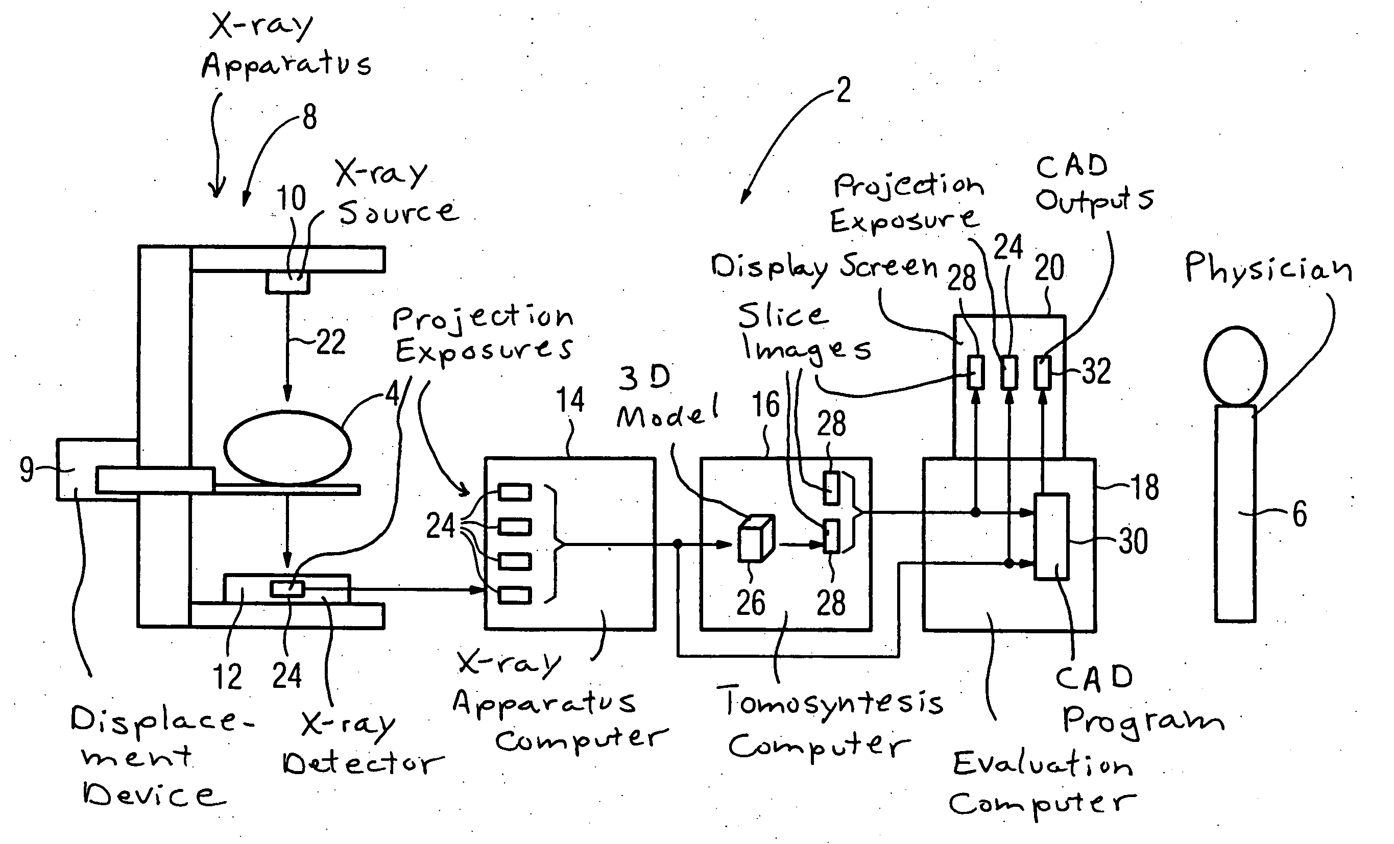
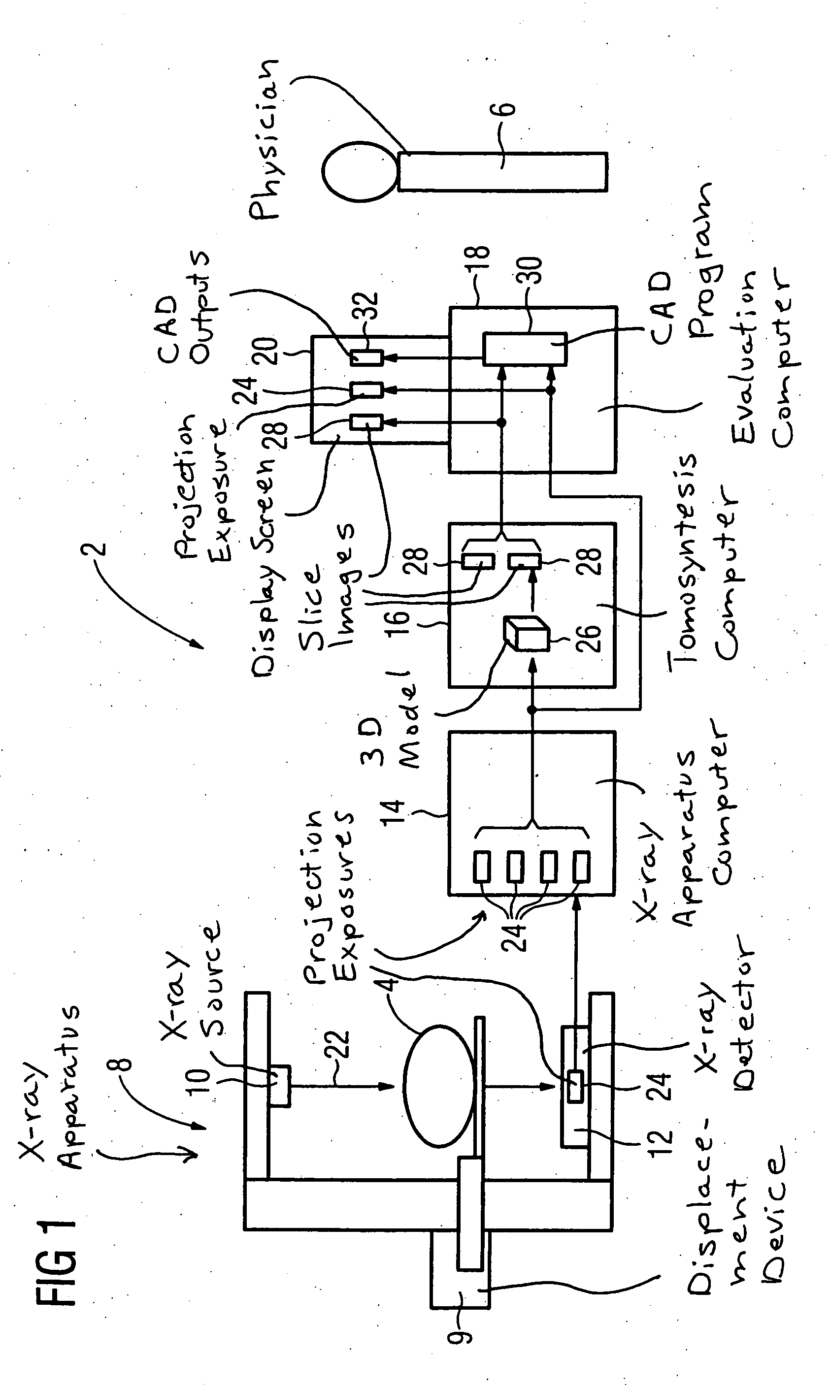
[0034]FIG. 1 shows a tomosynthesis device 2 for medical evaluation in three-dimensional imaging, i.e., in the framework of a tomosynthesis, with a patient 4 to be examined and a physician 6 conducting the examination. The tomosynthesis device 2 has an x-ray apparatus 8 with an x-ray source 10, a mechanical displacement device 9 and a digital x-ray detector 12 and a computer 14 belonging to the x-ray apparatus 8, a tomosynthesis computer 16 and a medical evaluation computer 18 with a screen 20.

[0035] The examination of the patient 4, for example the mammography of a female breast, is initiated at what is known as the acquisition workstation, in the form of the computer 14 which serves as a workstation for the medical personnel or the physician 6. The patient 4 is irradiated by x-rays 22 emanating from the x-ray source 10 and individual projections or projection exposures 24 are generated in the detector 12. The patient 4 and / or the x-ray source 10 and detector 12 are successively va...


PUM


Abstract

In a method for medical evaluation in three-dimensional imaging, in particular in mammography, projection exposures of a subject to be examined are generated and are stored in electronic form. Slice images are reconstructed from the projection exposures with a reconstruction method. A physician reviews and medically evaluates the slice images and marks a positive medical finding in the slice image with a first marker. A CAD system medically evaluates the slice images and marks a positive medical finding in the slice image with a second marker. A slice image with a first marker and a second marker deviating from one another is reconsidered and reevaluated by the physician.

Description

BACKGROUND OF THE INVENTION [0001] 1. Field of the Invention [0002] The present invention concerns a method for evaluation of medical findings in three-dimensional imaging, in particular in mammography. [0003] 2. Description of the Prior Art [0004] Imaging methods are of increasing importance in medical technology. The subject of the examination is a human or animal body. Classical methods such as, for example, the x-ray radiology, in which two-dimensional images of a body region to be examined are generated, have existed for a long time. In addition to this are modern methods, such as nuclear magnetic resonance tomography and computed tomography, which also generate three-dimensional images or, respectively, approximate, quasi-three-dimensional images (via slice representation). [0005] In digital tomosynthesis as is known, for example, from DE 198 42 944 A1, 3D information that is provided in the form of reconstructed slices is acquired with a digital projection x-ray system. [0006...

Claims


Application Information

Patent Timeline
Patent Type & Authority Applications(United States)
IPC IPC(8): A61B5/05
CPCA61B6/502G06T7/0012G06T2207/30068
Inventor MERTELMEIER, THOMAS
Owner SIEMENS AG
Why Eureka
Try Eureka