Insertion support system for specifying a location of interest as an arbitrary region and also appropriately setting a navigation leading to the specified region

a technology of insertion support and arbitrary region, which is applied in the field of insertion support system, can solve the problems of difficult to make the distal end of the endoscope correctly reach the target location within a short time period

Active Publication Date: 2006-08-31
OLYMPUS CORP
View PDF1 Cites 12 Cited by
  • Summary
  • Abstract
  • Description
  • Claims
  • Application Information

AI Technical Summary

Problems solved by technology

In a duct within the body that branches in multiple stages, such as the bronchi, if the abnormal area is located near a periphery of a branch, it is difficult to make the distal end of the endoscope correctly reach a target location within a short time period.

Method used


Image

  • Insertion support system for specifying a location of interest as an arbitrary region and also appropriately setting a navigation leading to the specified region
  • Insertion support system for specifying a location of interest as an arbitrary region and also appropriately setting a navigation leading to the specified region
  • Insertion support system for specifying a location of interest as an arbitrary region and also appropriately setting a navigation leading to the specified region

Examples


embodiment 1

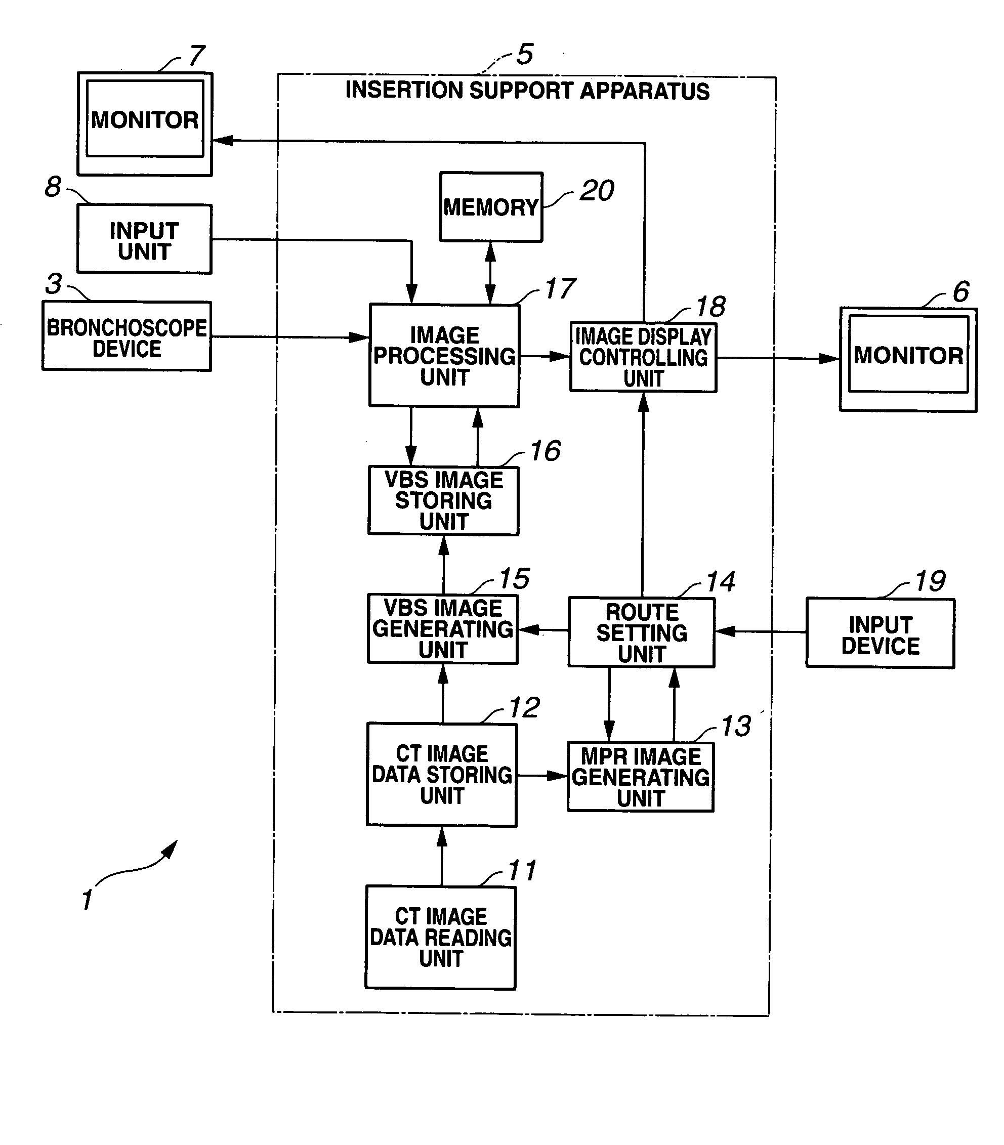
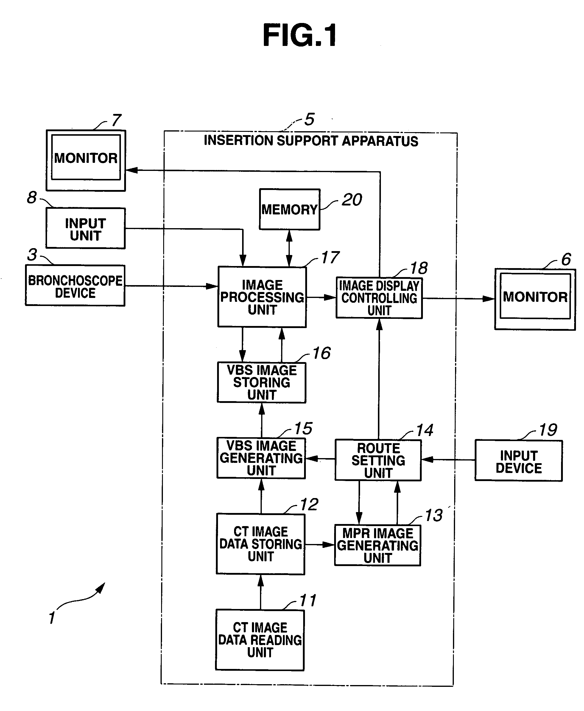
[0069] As illustrated in FIG. 1, a bronchi insertion support system 1 according to the present Embodiment 1 includes a bronchoscope device 3 and an insertion support apparatus 5.

[0070] The insertion support apparatus 5 supports insertion of the bronchoscope device 3 into the bronchi by generating a virtual endoscope image (hereinafter referred to as a VBS image) of the interior of the bronchi on the basis of CT image data, combining the VBS image with an endoscope image (hereinafter referred to as a live image) obtained by the bronchoscope device 3, and displaying a resultant image on a monitor 6.

[0071] The bronchoscope device 3 includes a bronchoscope having image picking-up means, a light source for supplying illuminating light to the bronchoscope, a camera controlling unit for performing signal processing on an image pickup signal sent by the bronchoscope, and the like, which are not illustrated in the figure. The bronchoscope device 3 inserts the bronchoscope into the bronchi ...

embodiment 2

[0104] As illustrated in FIG. 19, a bronchi insertion support system 301 according to the present Embodiment 2 includes a bronchoscope device 303 and an insertion support apparatus 305.

[0105] The insertion support apparatus 305 supports insertion of the bronchoscope device 303 into the bronchi by generating a virtual endoscope image (hereinafter referred to as a VBS image) of the interior of the bronchi on the basis of CT image data, combining the VBS image with an endoscope image (hereinafter referred to as a live image) obtained by the bronchoscope device 303, and displaying a resultant image on a monitor 306.

[0106] The bronchoscope device 303 includes a bronchoscope having image picking-up means, a light source for supplying illuminating light to the bronchoscope, a camera controlling unit for performing signal processing on an image pickup signal sent by the bronchoscope, and the like, which are not illustrated in the figure. The bronchoscope device 303 inserts the bronchoscop...

embodiment 3

[0146] As illustrated in FIG. 39, a bronchi insertion support system 501 according to the present Embodiment 3 includes a bronchoscope device 503 and an insertion support apparatus 505.

[0147] The insertion support apparatus 505 supports insertion of the bronchoscope device 503 into the bronchi by generating a virtual endoscope image (hereinafter referred to as a VBS image) of the interior of the bronchi on the basis of CT image data, combining the VBS image with an endoscope image (hereinafter referred to as a live image) obtained by the bronchoscope device 503, and displaying a resultant image on a monitor 506.

[0148] The bronchoscope device 503 includes a bronchoscope having image picking-up means, a light source for supplying illuminating light to the bronchoscope, a camera controlling unit for performing signal processing on an image pickup signal sent by the bronchoscope, and the like, which are not illustrated in the figure. The bronchoscope device 503 inserts the bronchoscop...


PUM


Abstract

According to an insertion support system of the present invention, when a biopsy area is specified at a periphery of the bronchi, the barycenter of the biopsy area is extracted. A circle centering on the barycenter is determined as a search area. The search area is expanded until the bronchi are located within the search area. A point in the search area to which the bronchi first reach is determined as an end point. A first route choice connecting the end point and a start point is determined. If the first route choice has not been registered yet, the first route choice is registered as a first registered route. Accordingly, a location of interest can be specified as an arbitrary region, and navigation leading to the specified region is appropriately set.

Description

CROSS REFERENCE TO RELATED APPLICATION [0001] This application is a continuation application of PCT / JP2004 / 016034 filed on Oct. 28, 2004 and claims the benefit of Japanese Applications No. 2003-373808 filed in Japan on Oct. 31, 2003, No. 2004-128489 filed in Japan on Apr. 23, 2004, and No. 2004-128490 filed in Japan on Apr. 23, 2004, the entire contents of each of which are incorporated herein by their reference.BACKGROUND OF THE INVENTION [0002] 1. Field of the Invention [0003] The present invention relates to an insertion support system for supporting insertion of an endoscope. [0004] 2. Description of the Related Art [0005] In recent years, diagnosis using an image has been widely performed. For example, a cross-sectional image of a subject is captured by using an X-ray CT (Computed Tomography) apparatus or the like to obtain three-dimensional image data of the subject. The obtained three-dimensional image data is then used for diagnosing a target area. [0006] The CT apparatus co...

Claims


Application Information

Patent Timeline
Patent Type & Authority Applications(United States)
IPC IPC(8): A61B5/05A61B1/00A61B6/00A61B19/00
CPCA61B1/2676A61B5/06A61B6/00A61B19/20A61B90/10
Inventor AKIMOTO, SHUNYAOHNISHI, JUNICHI
Owner OLYMPUS CORP
Why Eureka
Try Eureka