Method for imaging lesion and lesion imaging system

a technology for lesion imaging and lesion, applied in the field of patient imaging lesions, can solve the problems of difficult to accurately anticipate the contrast effectiveness based on experience alone, and achieve the effect of suitable contrast

Inactive Publication Date: 2006-10-19
MEDRAD INC.
View PDF2 Cites 16 Cited by
  • Summary
  • Abstract
  • Description
  • Claims
  • Application Information

AI Technical Summary

Benefits of technology

[0018] Thus, it is possible to obtain a more suitable contrast effect, which is necessary for diagnosis.

Problems solved by technology

The contrast effect that is obtained can vary according to infusion conditions such as the infusion rate, and according to the physical dimensions of the patient 6, however, and it is difficult to correctly anticipate the contrast effectiveness based on experience alone.

Method used


Image

  • Method for imaging lesion and lesion imaging system
  • Method for imaging lesion and lesion imaging system
  • Method for imaging lesion and lesion imaging system

Examples


Embodiment Construction

Overall Structure

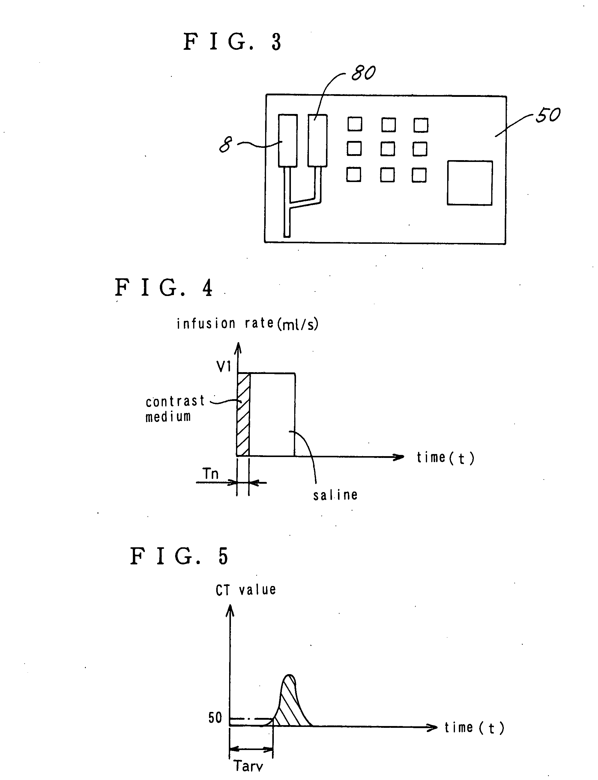
[0032] An example of the present invention is described in detail below using the drawings. This example is characterized in that simply by infusing a small amount of contrast medium in advance it is possible predict the contrast effect when an amount of contrast medium that is required for testing is infused.

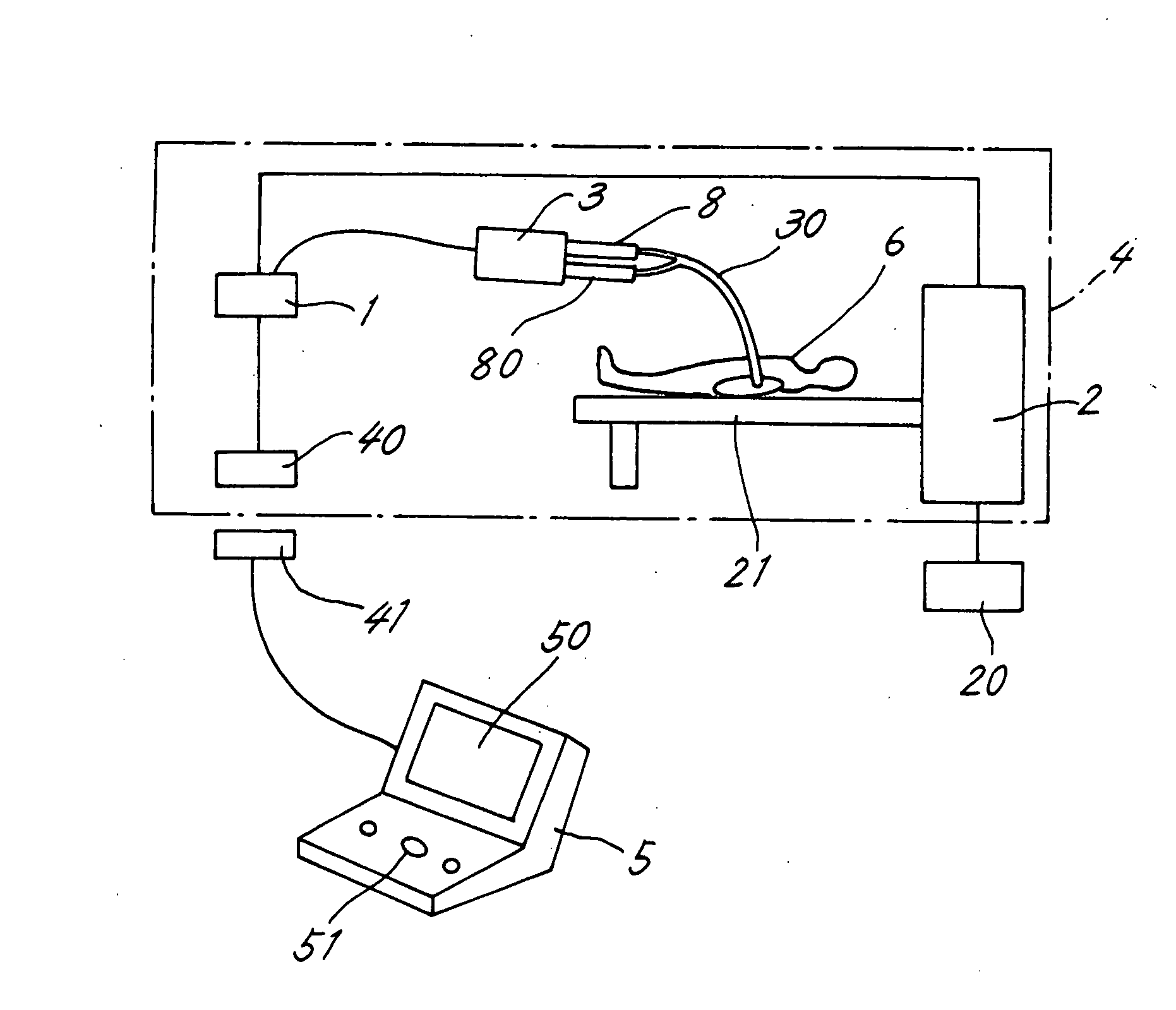
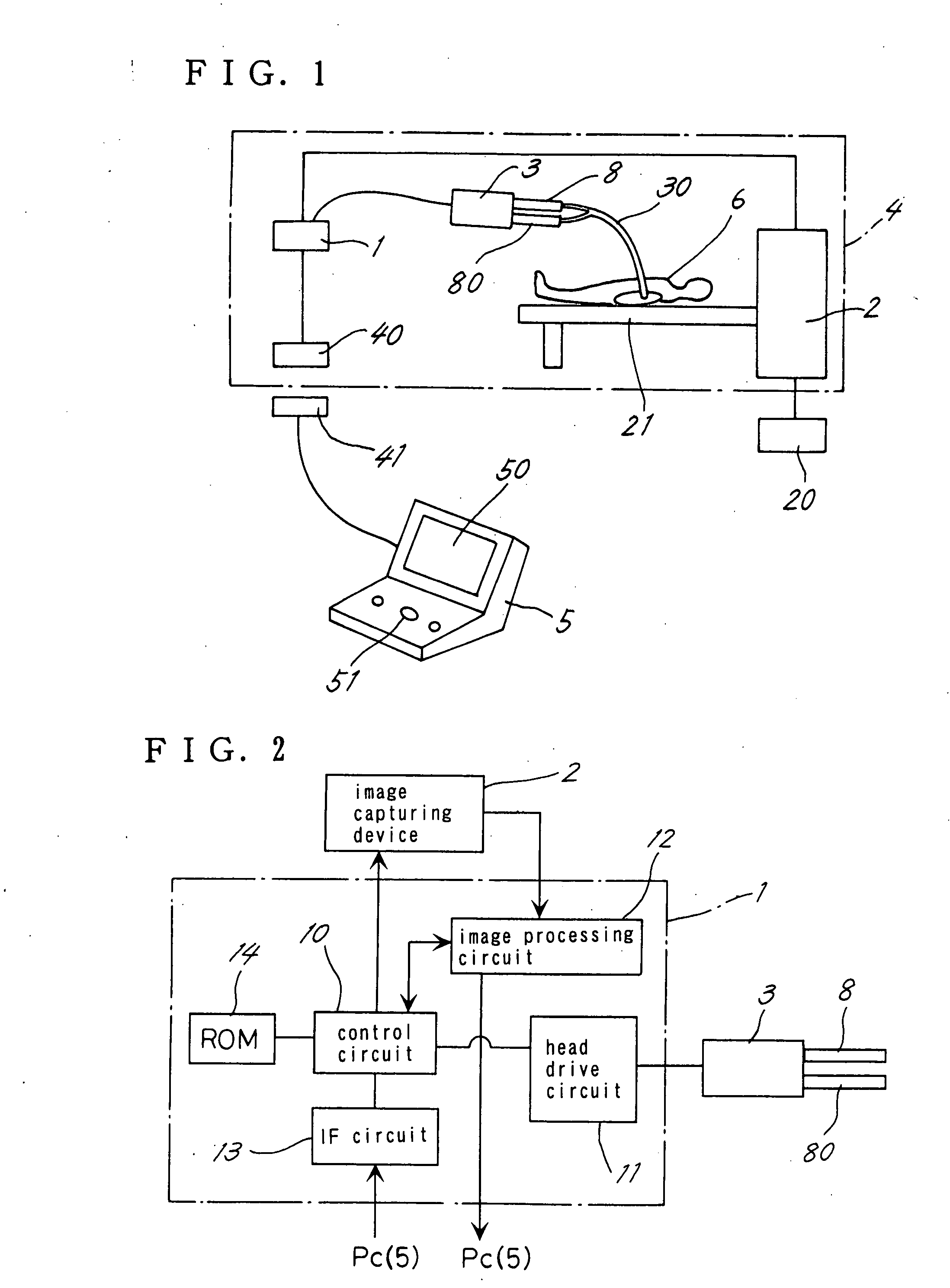
[0033]FIG. 1 is a block diagram of the lesion imaging system according to this example. An image capturing device 2, which is a CT device furnished with an injector head 3, a head control unit 1, a first infrared transceiver 40 and a display 20, is arranged inside an imaging room 4. A second infrared transceiver 41 and a PC 5 in serial communication with the first infrared transceiver 40 are arranged outside the imaging room 4. The PC 5 is provided with a screen 50 and an operation button 51, as is the case normally, and a physician or testing technician inputs the initial infusion conditions for the contrast medium, such as the infusion rate, through the PC...


PUM


Abstract

The invention provides a method for imaging a lesion area that includes a process of infusing a small amount of contrast medium from the syringe for a time Tn and using an image capturing device to measure a change in blood contrast medium concentration, a process of summing data that are based on the change in blood contrast medium concentration during the time Tn over a period of a required infusion time Tc for an amount of contrast medium necessary for diagnosis, to predict the contrast effect during the required infusion time Tc, and a process of displaying the predicted contrast effect and urging determination of whether or not to change the infusion conditions of the contrast medium.

Description

BACKGROUND OF THE INVENTION [0001] 1. Technical Field [0002] The present invention relates to methods for imaging lesions in a patient and lesion imaging systems for creating a diagnostic image of the patient. [0003] 2. Description of the Related Art [0004] To create diagnostic images of lesions, conventionally, a contrast medium is infused into a patient and then x-rays are irradiated onto the patient. The contrast medium in such cases is a pharmaceutical agent whose x-ray absorbency is different from that of human tissue, and for example is barium sulfate or an iodine solution. Of these, barium sulfate is administered orally, whereas iodine solutions are frequently used for injection or infusion into the body, but in the following description the contrast medium is not limited to iodine solutions. [0005]FIG. 10 is a block diagram of a lesion imaging system that has been proposed in the past (for example, see JP H07-204176A). This system is for controlling the infusion of contrast ...

Claims


Application Information

Patent Timeline
Patent Type & Authority Applications(United States)
IPC IPC(8): A61B6/00
CPCA61M5/007A61B6/481
Inventor KAWAMOTO, YASUSHI
Owner MEDRAD INC.
Why Eureka
Try Eureka