CT/MRI heart isolation using a graph cut algorithm

a graph cut and algorithm technology, applied in the field of graphical processing methods and systems, can solve the problems of not being work is not relevant for the instant disclosure purpose, and the segmentation provided by these prior art methods is generally slow, so as to achieve the effect of efficiently isolating the whole hear

Active Publication Date: 2006-10-26
SIEMENS MEDICAL SOLUTIONS USA INC
View PDF1 Cites 33 Cited by
  • Summary
  • Abstract
  • Description
  • Claims
  • Application Information

AI Technical Summary

Problems solved by technology

To assist cardiologists, techniques to segment the chambers of the heart for diagnostic purposes have been developed, but these techniques have not been used to visualize the heart surface.
A very large body of work exists related to the segmentation of the left ventricle of the heart in two-dimensional (2D) images, but this work is not relevant for purposes of the instant disclosure.
The segmentation provided by these prior art methods is generally slow because of the need to simulate mechanical deformations of a graphical model in 3D.
These methods are also inefficient at isolating the whole heart because the focus is on segmenting the chambers of the heart and not the surface of the heart.

Method used


Image

  • CT/MRI heart isolation using a graph cut algorithm
  • CT/MRI heart isolation using a graph cut algorithm
  • CT/MRI heart isolation using a graph cut algorithm

Examples


Embodiment Construction

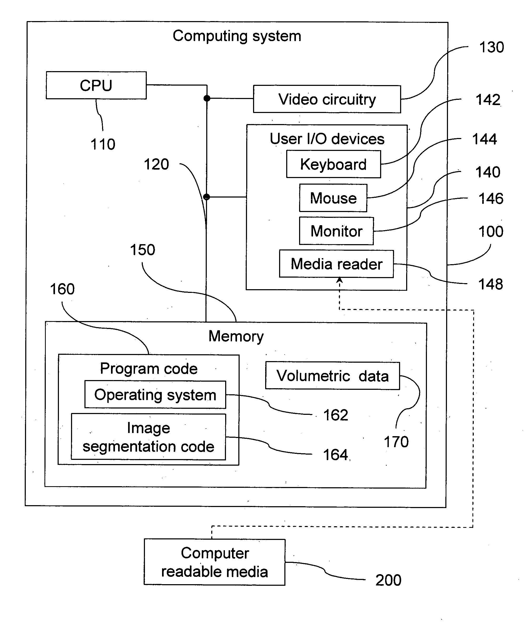
[0018] In accordance with an aspect of the present invention, volumetric data, as obtained from medical-imaging equipment, is processed to present to a user a visually useful image corresponding to the volumetric data. The volumetric data may be obtained, for example, from whole heart magnetic resonance angiography (MRA), or CT scans. It will be appreciated that volumetric data obtained by other means may also be utilized. That is, the present invention is not limited to specific types of volumetric data, file formats, voxel or pixel resolutions, or the like. The volumetric data may be thought of as describing a plurality of specific locations in space, each with a corresponding intensity value. Of course, the volumetric data may contain additional information, but such additional information is not required for the purposes of the following disclosure.

[0019] The present invention method may be implemented in the form of a computer program executable on any suitable computing devic...


PUM


Abstract

A method and related system for automatically and efficiently isolating the heart in Computer Tomography (CT) or Magnetic Resonance Imaging cardiac scans is disclosed. The method involves segmenting a heart within a set of volumetric data. In accordance with one aspect of the present invention, the set of volumetric data is processed to determine the minimum value of an energy function having a first term, a second term and a third term. The heart is segmented based on the processing of the set of volumetric data.

Description

CROSS-REFERENCE TO RELATED APPLICATIONS [0001] This application claims the benefit of U.S. Provisional Application No. 60 / 665,893, filed Mar. 28, 2005, which is incorporated herein by reference.BACKGROUND OF THE INVENTION [0002] The present invention relates to graphical processing methods and systems. More particularly, graphical methods and related systems relating to the segmenting of an organ, such as a heart, from medical imaging information are disclosed. [0003] Cardiac Computer Tomography (CT) is a fast, non-invasive, sub-millimeter resolution medical imaging tool. However, three-dimensional (3D) visualization requires human interaction to prepare the data, and to remove structures that do not belong to the heart (such as the lungs, liver, ribs, etc.). To assist cardiologists, techniques to segment the chambers of the heart for diagnostic purposes have been developed, but these techniques have not been used to visualize the heart surface. For these methods to be suitable acro...

Claims


Application Information

Patent Timeline
Patent Type & Authority Applications(United States)
IPC IPC(8): G06K9/34G06V10/28
CPCG06K9/38G06T7/0083G06T2207/30048G06T2207/10081G06T2207/10088G06T7/0089G06T7/12G06T7/149G06V10/28
Inventor FLORIN, CHARLESMOREAU-GOBARD, ROMAINBOYKOV, YURIFUNKA-LEA, GARETH
Owner SIEMENS MEDICAL SOLUTIONS USA INC
Why Eureka
Try Eureka