
Micromachined tissue anchors for securing implants without sutures

a micro-machined, tissue anchor technology, applied in the field of tissue anchoring systems and methods, can solve the problems of invasive and potentially damaging suturing devices to the iris, invasive suturing and other securing techniques, and potentially damaging the surrounding tissue, and achieve the effect of safe and practical way

Inactive Publication Date: 2006-11-02
CALIFORNIA INST OF TECH
View PDF9 Cites 26 Cited by
  • Summary
  • Abstract
  • Description
  • Claims
  • Application Information

AI Technical Summary

Benefits of technology

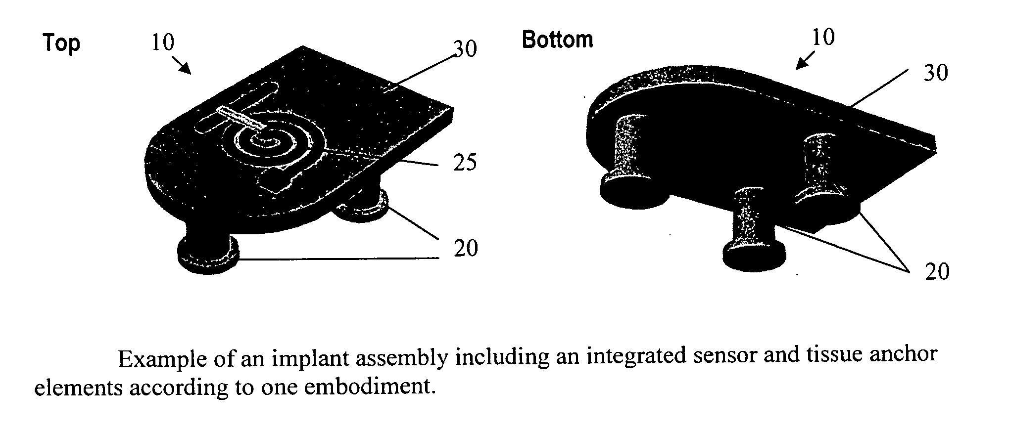
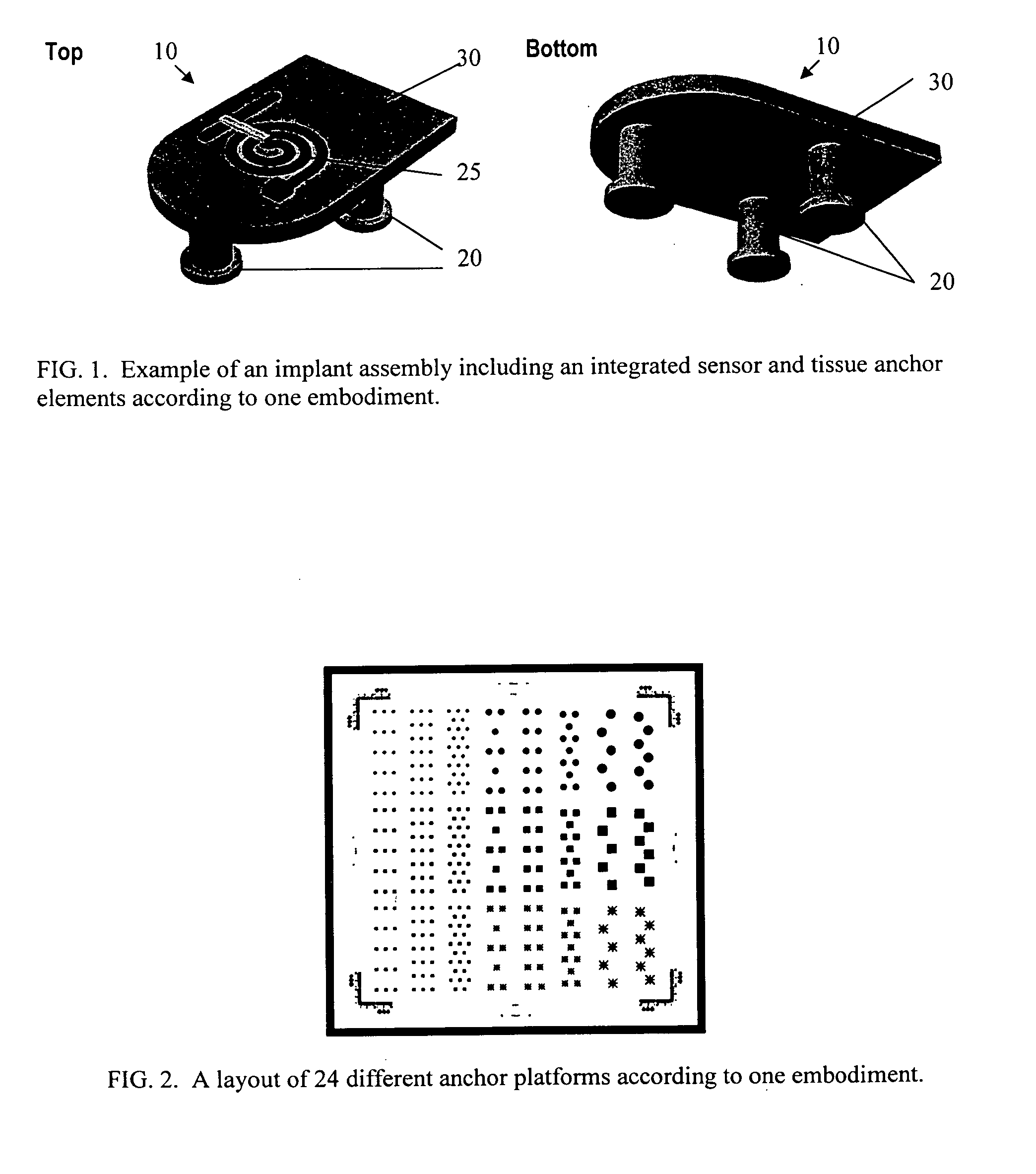
The present invention provides systems and methods for attaching an implant device to tissue by mechanically (and non-invasively) anchoring the device to the tissue. The anchor structures on the implant device match the topology and features of the tissue where the implant is to occur, making it secure and practical to attach an implant device in a non-invasive way. The implant device can include sensing elements and other features, and the method includes fabricating the anchor structures and securing the device to tissue. The technical effects include a safer and less invasive way to attach an implant device to tissue.

Problems solved by technology

However, conventional methods of suturing the device to the iris are invasive and potentially damaging.
Similarly, for implant devices designed for other tissue locations, suturing and other securing techniques are invasive and potentially damaging to surrounding tissue.

Method used


Image

  • Micromachined tissue anchors for securing implants without sutures
  • Micromachined tissue anchors for securing implants without sutures
  • Micromachined tissue anchors for securing implants without sutures

Examples


examples

[0039] Two generations of prototype intraocular implant devices similar to those described herein were implanted into rabbits and are being evaluated for adaptation to humans. In both versions of the anchors, the act of resting the anchors on top of a rabbit's iris was enough to hold the device in place. Although the precise removal force was not quantitatively determined, the mechanical locking of the anchors with the iris was more than sufficient to keep the devices secured to the iris. A significant amount of force is necessary to remove the devices once in place (such forces are greater than that exerted on the device during normal eye movement). FIG. 13 is a picture illustrating a device with anchors anchored on human skin. The device remains secured to the finger tissue even during serious shaking of the finger.


PUM


Abstract

Systems and methods for attaching an implant device to tissue by mechanically (and non-invasively) anchoring the device to the tissue. The systems and methods provide a safe, practical way to attach an implant device to tissue in a non-invasive, or less invasive manner. According to the present invention, an implant device includes one or more protruding anchor-like structures for securely attaching to tissue. One or more device features, such as sensing elements, may be incorporated on the implant device. The anchor structures are configured and arranged to match the topology and features of the tissue environment where implant is to occur.

Description

CROSS-REFERENCES TO RELATED APPLICATIONS [0001] This application claims the benefit of U.S. Provisional Application No. 60 / 659,520 (Attorney docket No. 020859-008300US; Client Ref. CIT-4325-P), filed Mar. 8, 2005, the disclosure of which is incorporated herein by reference in its entirety.STATEMENT AS TO RIGHTS TO INVENTIONS MADE UNDER FEDERALLY SPONSORED RESEARCH OR DEVELOPMENT [0002] The government may have certain rights to the invention based on National Science Foundation Grant EEC-0310723.BACKGROUND OF THE INVENTION [0003] The present invention relates generally to systems and methods for securing implants to tissue, and more particularly to implant structures and devices with tissue anchors for use in securing the implant structures to tissue without sutures. [0004] Biomedical implants often require a means of attaching to the body in order to secure the device in the desired location. One such device might be an implantable passive sensor for determining intraocular pressure...

Claims


Application Information

Patent Timeline
Patent Type & Authority Applications(United States)
IPC IPC(8): A61F2/02A61B3/16
CPCA61B3/16A61B5/6821A61B5/6882
Inventor MENG, ELLISTAI, YU-CHONGRODGER, DAMIEN C.CHEN, PO-JUIHUMAYUN, MARK
Owner CALIFORNIA INST OF TECH
Learn More