X-ray diagnostic apparatus, imaging angle determination device, program storage medium, and method

a diagnostic apparatus and imaging angle technology, applied in the field of x-ray diagnostic apparatus, imaging angle determination device, program storage medium, and method, can solve the problems of difficult automation, hindering the flow of examination, and inability to perform proper measuremen

Active Publication Date: 2007-02-01
TOSHIBA MEDICAL SYST CORP
View PDF0 Cites 17 Cited by
  • Summary
  • Abstract
  • Description
  • Claims
  • Application Information

AI Technical Summary

Benefits of technology

[0010] An X-ray diagnostic apparatus according to the present invention comprises an X-ray tube, an X-ray detector, a support mechanism which movably supports the X-ray tube and the X-ray detector, a storage unit which stores data of a three-dimensional model associated with a standard blood vessel str

Problems solved by technology

This makes it impossible to perform proper measurement.
In the former method, it is necessary to associate corresponding positions between frames, and it is difficult to automate this.
This operation always requires a doctor or technologist with rich clinical knowledge to perform designation, and henc

Method used


Image

  • X-ray diagnostic apparatus, imaging angle determination device, program storage medium, and method
  • X-ray diagnostic apparatus, imaging angle determination device, program storage medium, and method
  • X-ray diagnostic apparatus, imaging angle determination device, program storage medium, and method

Examples


Embodiment Construction

[0024] An embodiment of the present invention will be described below with reference to the views of the accompanying drawing. Note that this embodiment will exemplify an X-ray diagnostic apparatus having an imaging angle optimization support function, but may be provided as an imaging angle optimization support device which serves as part of an X-ray diagnostic apparatus. In addition, the embodiment can be provided as a program for causing a computer to implement processing for supporting the optimization of an imaging angle, and a storage medium storing the program.

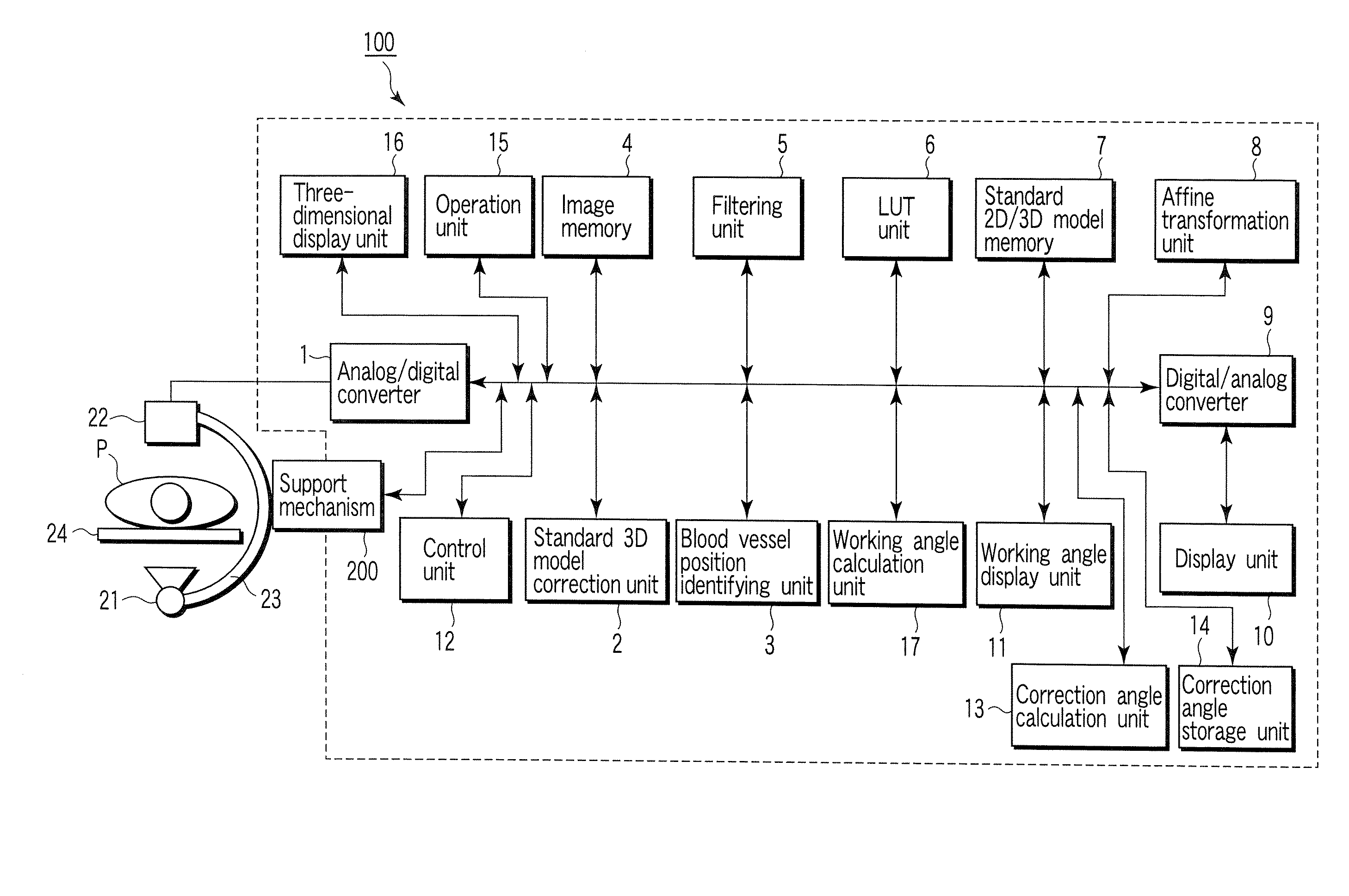
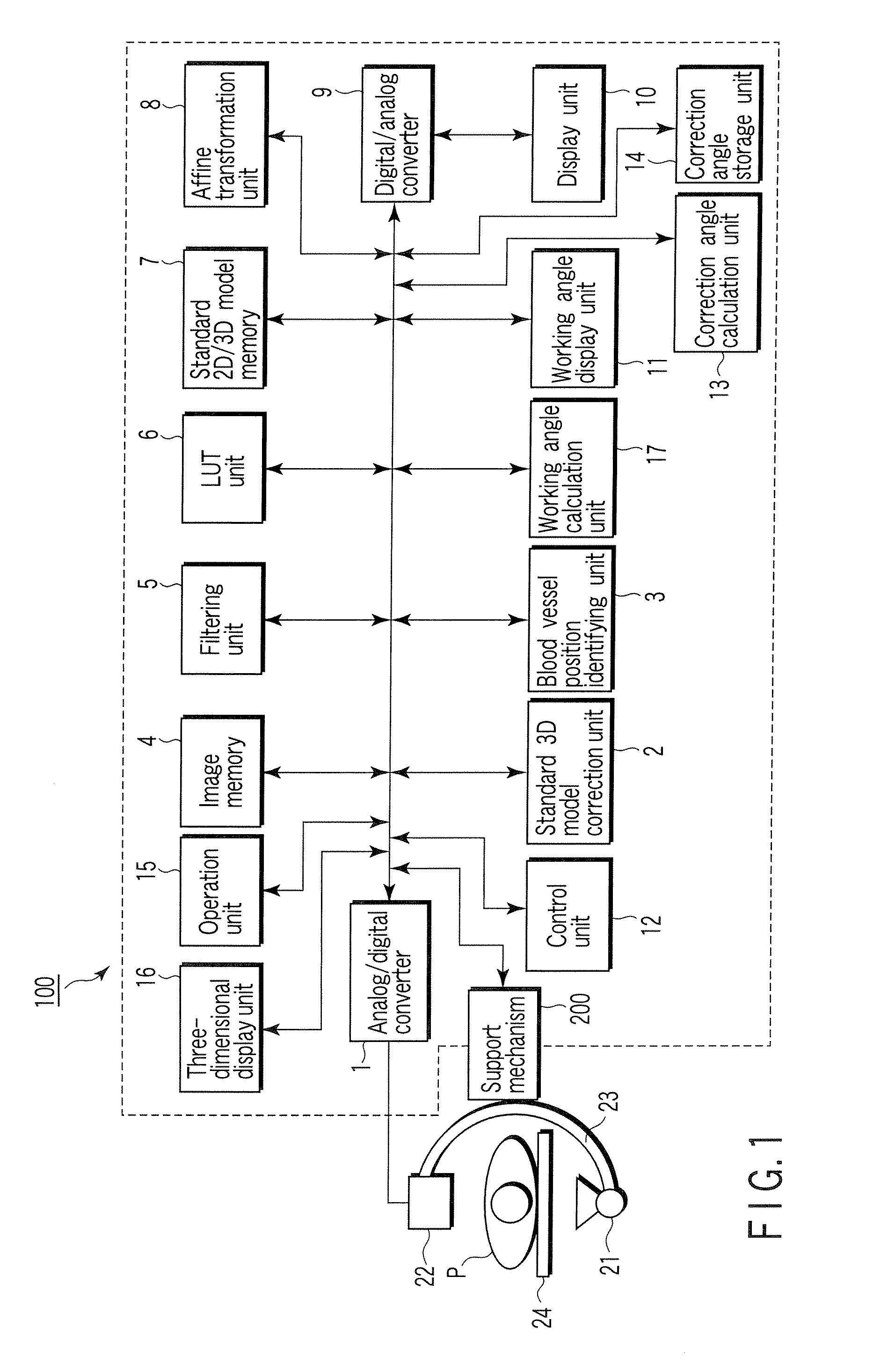
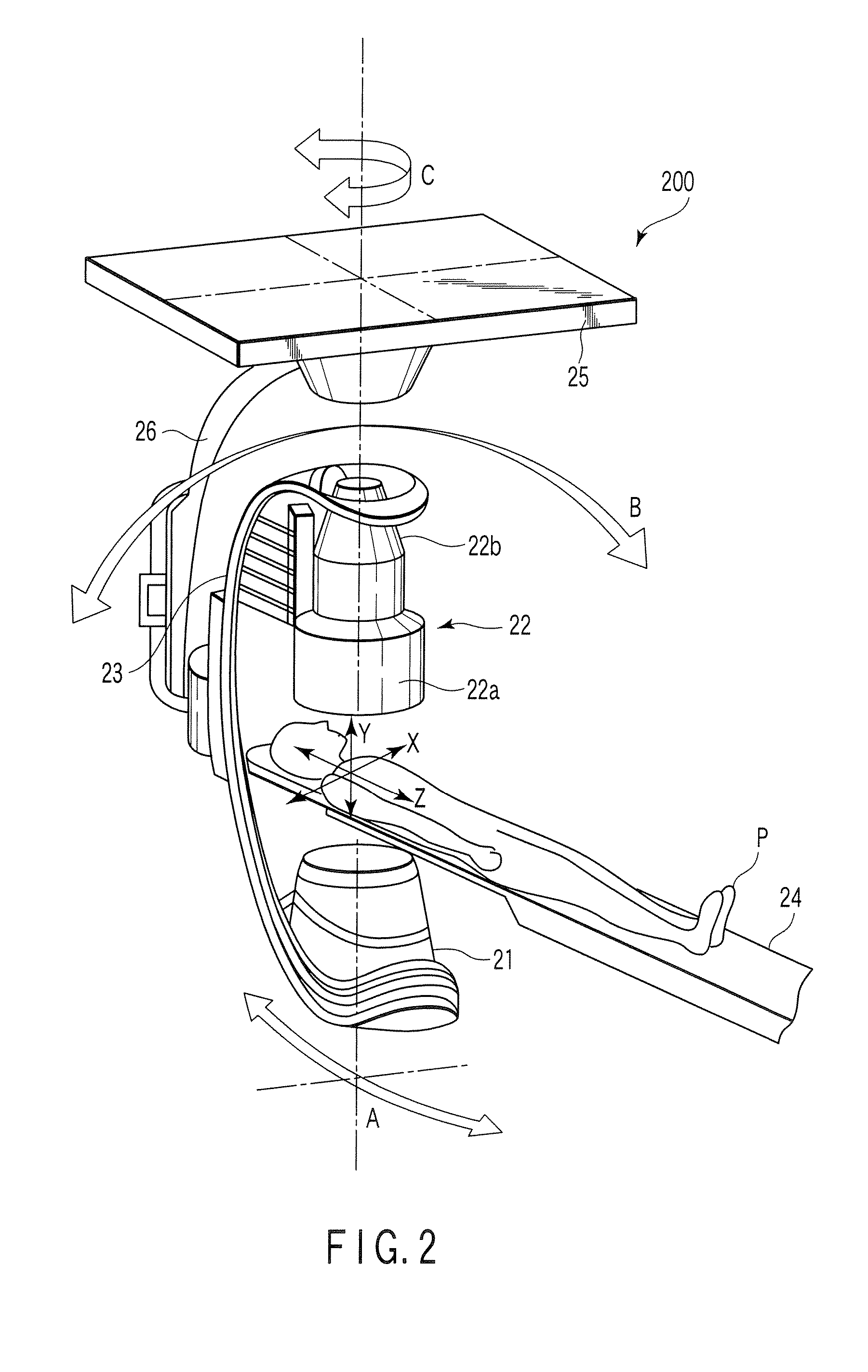
[0025]FIG. 1 shows the arrangement of the main part of the X-ray diagnostic apparatus according to this embodiment. An X-ray tube 21 generates X-rays upon reception of a high voltage (tube voltage) and filament current from a high voltage generator (not shown). The X-ray tube 21 is mounted on one end of a C-arm 23. An X-ray detector 22 is mounted on the other end of the C-arm 23. The X-ray detector 22 faces the X-ray t...


PUM


Abstract

An X-ray diagnostic apparatus includes an X-ray tube, an X-ray detector, a support mechanism which movably supports the X-ray tube and the X-ray detector, a storage unit which stores data of a three-dimensional model associated with a standard blood vessel structure, and a control unit which controls the support mechanism on the basis of the three-dimensional model so as to make an imaging central line between the X-ray tube and the X-ray detector become substantially orthogonal to a blood vessel axis of the three-dimensional model at a designated position.

Description

CROSS-REFERENCE TO RELATED APPLICATIONS [0001] This application is based upon and claims the benefit of priority from prior Japanese Patent Application No. 2005-207688, filed Jul. 15, 2005, the entire contents of which are incorporated herein by reference. BACKGROUND OF THE INVENTION [0002] 1. Field of the Invention [0003] The present invention relates to an X-ray diagnostic apparatus which has a high degree of freedom concerning an imaging angle owing to a mechanism including a C-arm and the like, an imaging angle determination device, a program storage medium, and a method. [0004] 2. Description of the Related Art [0005] In diagnosis using an X-ray diagnostic apparatus, and more specifically, in diagnosis of a case of stricture, imaging needs to be performed such that the axial direction of a blood vessel does not become parallel to the depth direction (projection direction) of X-rays. Note that angles of the imaging angles of X-rays which are used for medical treatment and diagno...

Claims


Application Information

Patent Timeline
IPC IPC(8): G01N23/04
CPCA61B6/4441A61B6/463A61B6/465A61B6/583A61B6/481A61B6/504A61B6/469
Inventor OHISHI, SATORU
Owner TOSHIBA MEDICAL SYST CORP
Why Eureka
Try Eureka