
Tissue culture microscope apparatus

a microscope and tissue technology, applied in the field of tissue culture microscopes, can solve the problems of out of focus objects, difference in temperature between specimens and reagents, and inability to properly observe specimens in the laboratory environment,

Inactive Publication Date: 2007-03-22
OLYMPUS CORP
View PDF10 Cites 23 Cited by
  • Summary
  • Abstract
  • Description
  • Claims
  • Application Information

AI Technical Summary

Problems solved by technology

The normal indoor environment in which a light microscope is used adversely affects the cell in culture because the cell environment in culture differs from such indoor environment.
Since an environment suitable for cell culture is maintained within the incubator (cell culture space), dosing through an opening of the incubator results in changes in the cell culture environment and causes a difference in temperature between the specimen and the reagent.
As a result, the dosing causes out of focus objects and has temperature effects on the cell.
However, since frequency of use of the reagent is low, for example, one dosing per experiment, such a specific dosing apparatus provides a low cost-effectiveness.
Also, installing the specific dosing apparatus makes the whole incubator complicated and thus makes it hard to clean.
Insufficient cleaning makes it easy to get mold.

Method used


Image

  • Tissue culture microscope apparatus
  • Tissue culture microscope apparatus
  • Tissue culture microscope apparatus

Examples


Embodiment Construction

[0018] Exemplary embodiments of the present invention will be described below with reference to the accompanying drawings.

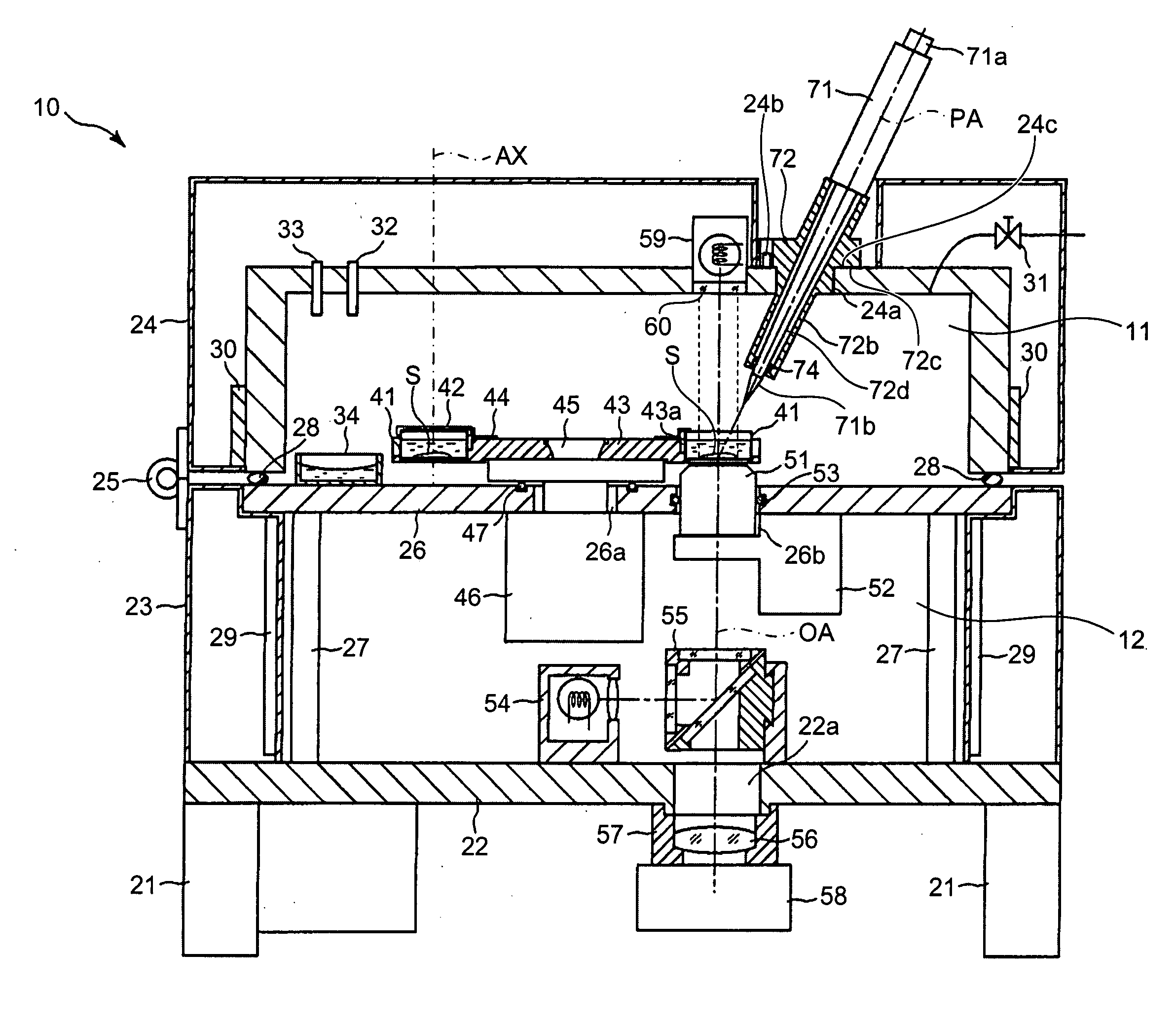
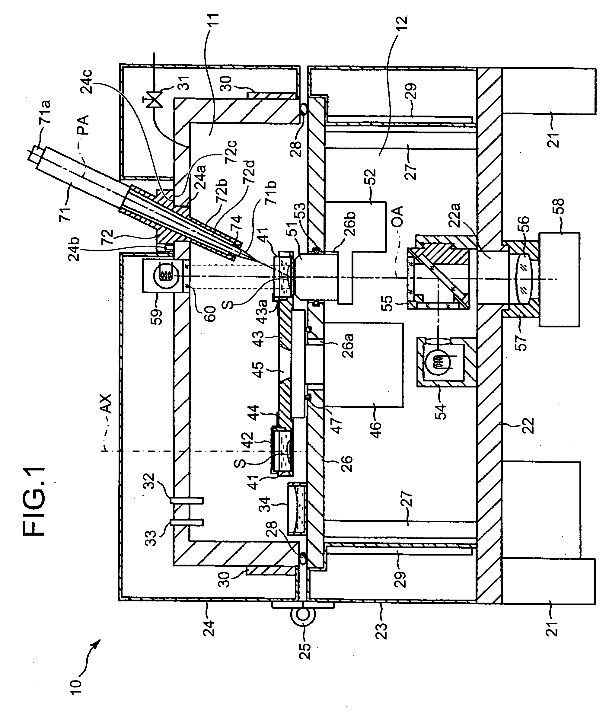
[0019]FIG. 1 is a cross-sectional view of a tissue culture microscope apparatus in accordance with the present invention. The tissue culture microscope apparatus mainly includes a culture unit allowing control of temperature, humidity, CO2 concentration of a specimen containing a cultured cell; and a microscope allowing enlarged observation of the specimen.

[0020] As shown in FIG. 1, the tissue culture microscope apparatus 10 includes a base member 22 supported by feet 21; a closed side wall 23 on the periphery of the base member 23; a separator 26 closing an upper opening of the side wall 23; and an opening / closing cover 24 having a open bottom.

[0021] The separator 26 is supported by a plurality of support posts 27 stood on the base member 22. The side wall 23 has a cavity serving as a thermal insulation space, and includes a heater 29 in the cavity.

[0022] Th...


PUM


Abstract

A tissue culture microscope apparatus includes a culture unit that includes a chamber in which a specimen is put, and maintains the chamber at a predetermined temperature to culture the specimen; an observation unit that forms an observation image of the specimen put in the chamber; and a liquid supply unit that stores a liquid in a protrusion portion penetrating into a wall of the chamber and protruding to an inside of the chamber, matches a temperature of the liquid with the temperature of the chamber, and injects the liquid from the protrusion portion to the specimen.

Description

CROSS-REFERENCE TO RELATED APPLICATIONS [0001] This application is based upon and claims the benefit of priority from Japanese Patent Applications No. 2005-276205, filed Sep. 22, 2005; and No. 2006-208876, filed Jul. 31, 2006, the entire contents of both of which are incorporated herein by reference. BACKGROUND OF THE INVENTION [0002] 1. Field of the Invention [0003] The present invention relates to a tissue culture microscope apparatus that allows observation of a cell in culture. [0004] 2. Description of the Related Art [0005] There has been a trend for molecular biological or biophysical research to use a cultured cell. The normal indoor environment in which a light microscope is used adversely affects the cell in culture because the cell environment in culture differs from such indoor environment. [0006] A specific tissue cell microscope is reported in which a light microscope and an incubator for maintaining an environment suitable for cell culture are combined together, allowi...

Claims


Application Information

Patent Timeline
IPC IPC(8): C12M1/34G01N21/01
CPCC12M23/48C12M41/14G02B21/30C12M41/46G01N2035/00425C12M41/36Y10S435/809
Inventor HASEGAWA, KAZUHIROKAGAYAMA, AKITSUGUTSUCHIYA, ATSUHIROHIRANO, RYUICHIKOYAMA, KENICHIENDO, HIDEAKI
Owner OLYMPUS CORP
Learn More