Automatic bone detection in MRI images

a bone detection and image technology, applied in image analysis, image enhancement, instruments, etc., can solve the problems of difficult automatic segmentation and classification of tissues in mri images, difficult design of automatic segmentation tools, and less effective mri than x-rays or ct for detecting destruction of bone structur

Inactive Publication Date: 2007-04-12
SIEMENS MEDICAL SOLUTIONS USA INC
View PDF7 Cites 38 Cited by
  • Summary
  • Abstract
  • Description
  • Claims
  • Application Information

AI Technical Summary

Benefits of technology

[0008] According to an exemplary embodiment of the present invention, a method is provided for detecting bone in magnetic resonance imaging (MRI) images. The method includes detecting bone borders using dark bone border intensity information from an MRI image, wherein dark bone border intensity information comprises low or zero signal intensity in MRI images. According to an exemplary embodiment of the present invention, a method is provided for detecting bone and bone disease using MRI images. The method includes: detecting and segmenting bone borders using dark bone border intensity information from an MRI image; and detecting bone disease within a segmented image region.

Problems solved by technology

While MRI offers excellent tissue discrimination and can provide diagnostic quality images, automatic segmentation and classification of tissues in MRI images is difficult due to intra- and inter-scan intensity inhomogeneities.
In addition to the high intensity variability within a single tissue type, in MRI images, several tissue types appear to have similar intensity and textural properties, making it harder to design automatic segmentation tools.
Although bone marrow lesions may be more accurately assessed by MRI than by other imaging modalities, MRI is less effective than X-rays or CT for detecting destruction of bone structure, because cortical bone does not produce a signal and appears black on T1- and T2-weighted sequences.

Method used


Image

  • Automatic bone detection in MRI images
  • Automatic bone detection in MRI images
  • Automatic bone detection in MRI images

Examples


Embodiment Construction

[0018] Hereinafter, exemplary embodiments of the present invention will be described in detail with reference to the accompanying drawings.

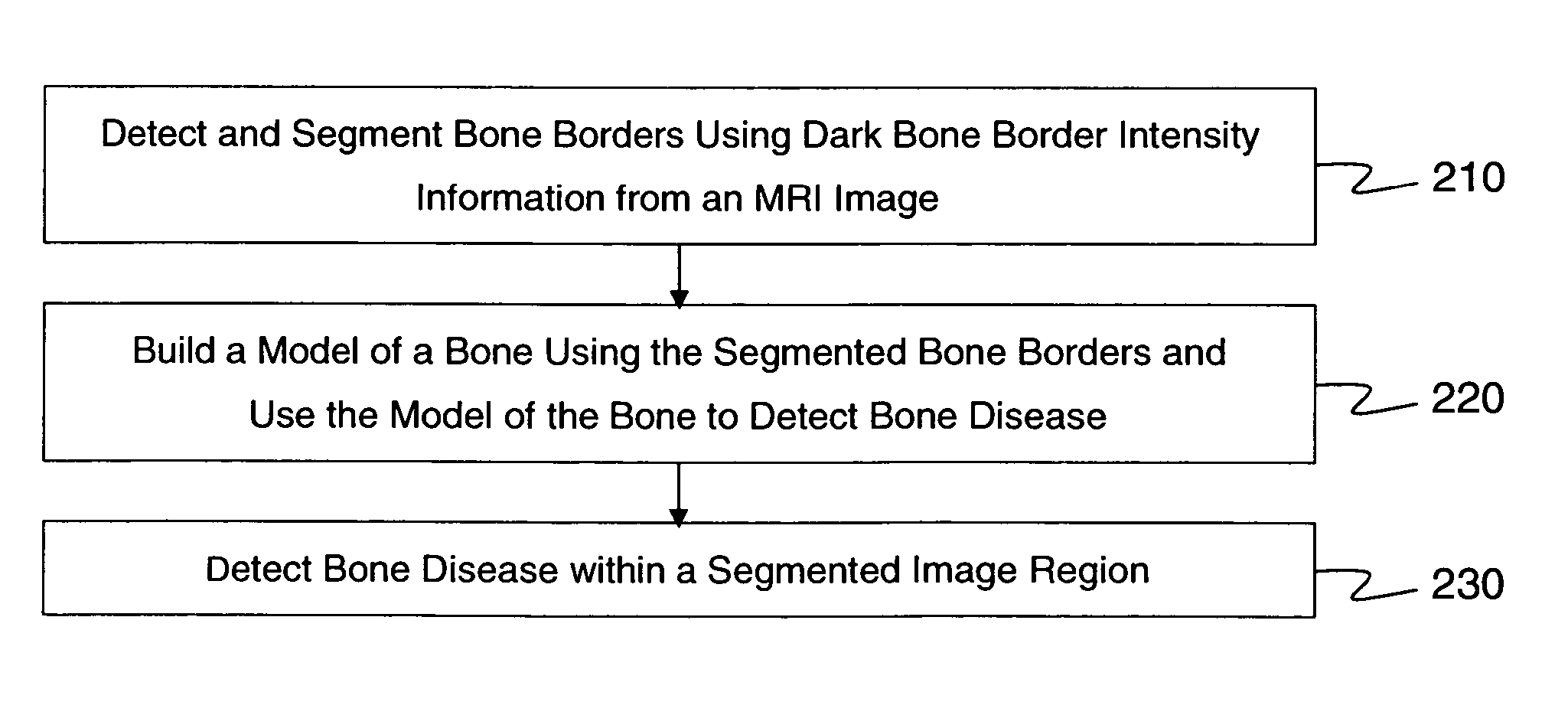
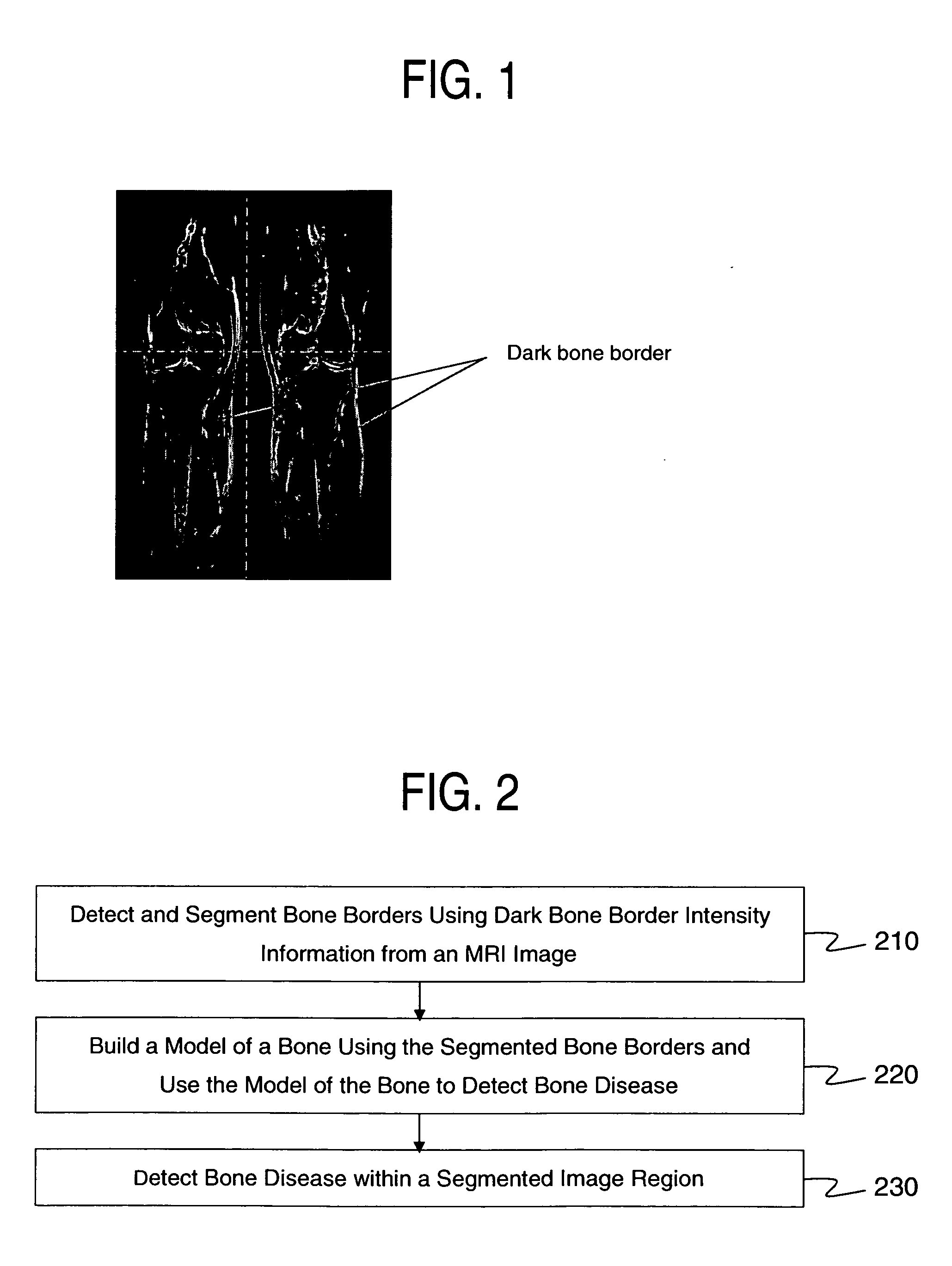
[0019] In exemplary embodiments of the present invention, black border around bones in magnetic resonance imaging (MRI) images is extracted and used to represent bone contour. For example, cortical bone and periosteum appear consistently dark on HASTE, T1 and T2 STIR (short tau-inversion recovery) MRI images. Intensity normalization can be used as a preprocessing step.

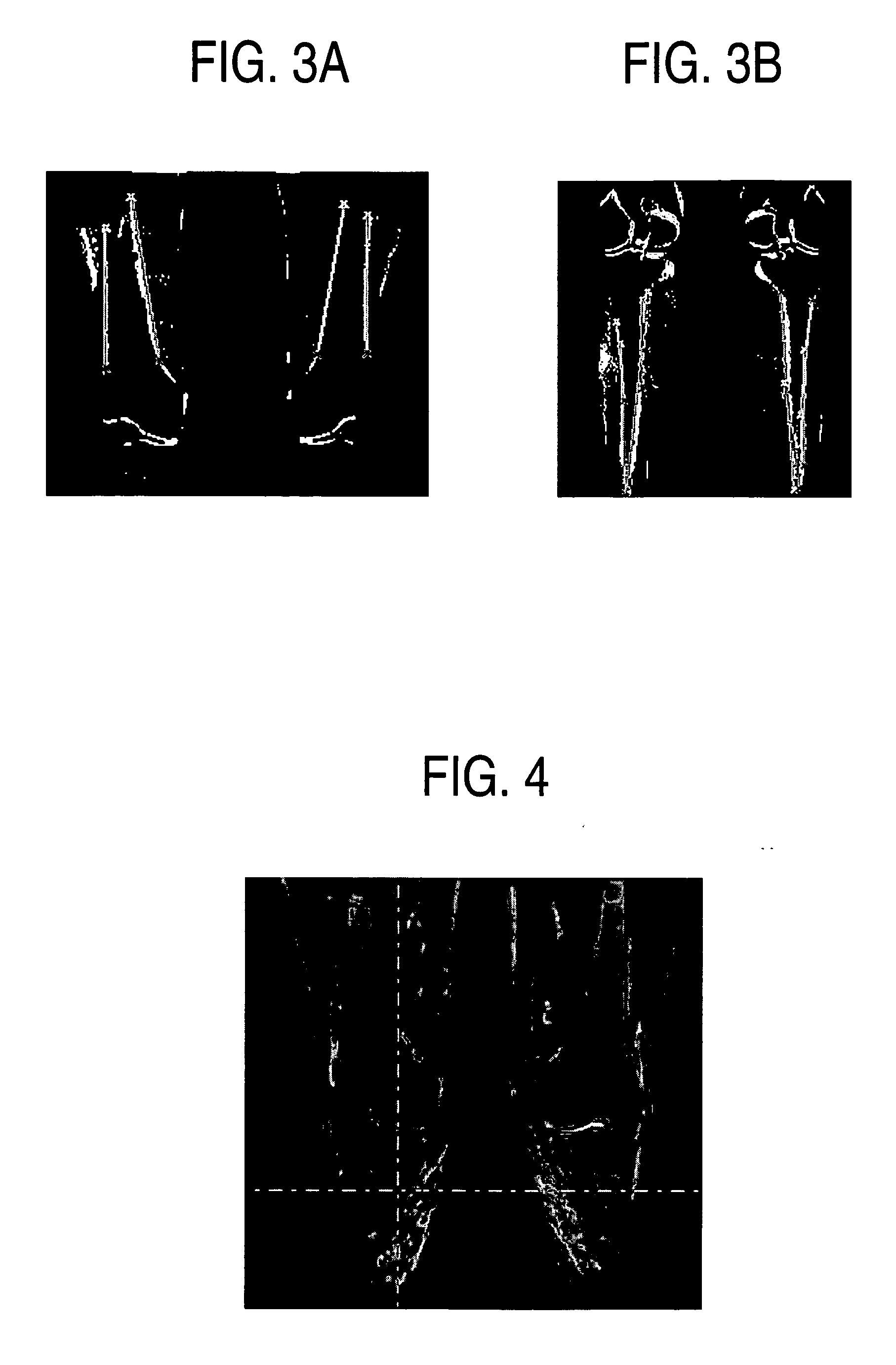
[0020] After the bone border is extracted, bone metastases can be detected as dark spots inside the bone, such as in the case of a T1 pulse sequence, or as bright spots, in the case of a T2 STIR pulse sequence. Further classification step using additional features may be included to automatically detect bone metastases sites and / or to help to discriminate them from false positive findings.

[0021] In a method of detecting bone in magnetic resonance imaging (MRI) images, according t...


PUM


Abstract

A method for detecting bone and bone disease using MRI images includes: detecting and segmenting bone borders using dark bone border intensity information from an MRI image; and detecting bone disease within a segmented image region.

Description

CROSS-REFERENCE TO RELATED APPLICATION [0001] This application claims the benefit of U.S. Provisional Application Ser. No. 60 / 724,391 (Attorney Docket No. 2005P18156US), filed Oct. 7, 2005 and entitled “Method and Apparatus for Automatic Bone and Bone Metastasis Detection in MRI Images,” the content of which is herein incorporated by reference in its entirety.BACKGROUND OF THE INVENTION [0002] 1. Technical Field [0003] The present disclosure relates to systems and methods for automatic bone detection in MRI images. [0004] 2. Discussion of Related Art [0005] In recent years, medical imaging has experienced an explosive growth due to advances in imaging modalities such as X-rays, computed tomography (CT), magnetic resonance imaging (MRI) and ultrasound. MRI has demonstrated a high potential in bone metastasis identification at an early stage of growth. While MRI offers excellent tissue discrimination and can provide diagnostic quality images, automatic segmentation and classification ...

Claims


Application Information

Patent Timeline
Patent Type & Authority Applications(United States)
IPC IPC(8): G06K9/00
CPCG06T7/0012G06T7/0083G06T7/0087G06T2207/10088G06T2207/20061G06T2207/30008G06T7/12G06T7/143
Inventor JEREBKO, ANNA
Owner SIEMENS MEDICAL SOLUTIONS USA INC
Why Eureka
Try Eureka